Explore
 Validate
 Learn
Learn Western blot
 Immunocytochemistry
Antibody data
- Antibody Data
- Antigen structure
- References [2]
- Comments [0]
- Validations
- Immunocytochemistry [1]
- Immunohistochemistry [1]
Submit
Validation data
Reference
Comment
Report error
- Product number
- HPA043922 - Provider product page

- Provider
- Atlas Antibodies
- Proper citation
- Atlas Antibodies Cat#HPA043922, RRID:AB_2678732
- Product name
- Anti-PC
- Antibody type
- Polyclonal
- Description
- Polyclonal Antibody against Human PC, Gene description: pyruvate carboxylase, Alternative Gene Names: PCB, Validated applications: ICC, IHC, WB, Uniprot ID: P11498, Storage: Store at +4°C for short term storage. Long time storage is recommended at -20°C.
- Reactivity
- Human
- Host
- Rabbit
- Conjugate
- Unconjugated
- Isotype
- IgG
- Vial size
- 100 µl
- Concentration
- 0.1 mg/ml
- Storage
- Store at +4°C for short term storage. Long time storage is recommended at -20°C.
- Handling
- The antibody solution should be gently mixed before use.
Submitted references Hypoxia-mediated repression of pyruvate carboxylase drives immunosuppression
Pyruvate carboxylation enables growth of SDH-deficient cells by supporting aspartate biosynthesis
Coleman M, Cotul E, Pfeil A, Devericks E, Safdar M, Monteiro M, Chen H, Ho A, Attaar N, Malian H, Kiesel V, Ramos A, Smith M, Panchal H, Mailloux A, Teegarden D, Hursting S, Wendt M
Breast Cancer Research 2024;26(1)
Breast Cancer Research 2024;26(1)
Pyruvate carboxylation enables growth of SDH-deficient cells by supporting aspartate biosynthesis
Cardaci S, Zheng L, MacKay G, van den Broek N, MacKenzie E, Nixon C, Stevenson D, Tumanov S, Bulusu V, Kamphorst J, Vazquez A, Fleming S, Schiavi F, Kalna G, Blyth K, Strathdee D, Gottlieb E
Nature Cell Biology 2015;17(10):1317-1326
Nature Cell Biology 2015;17(10):1317-1326
No comments: Submit comment
Supportive validation
- Submitted by
- Atlas Antibodies (provider)
- Main image

- Experimental details
- Immunofluorescent staining of human cell line A549 shows localization to mitochondria.
- Sample type
- Human

Supportive validation
- Submitted by
- Atlas Antibodies (provider)
- Enhanced method
- Orthogonal validation
- Main image
- Experimental details
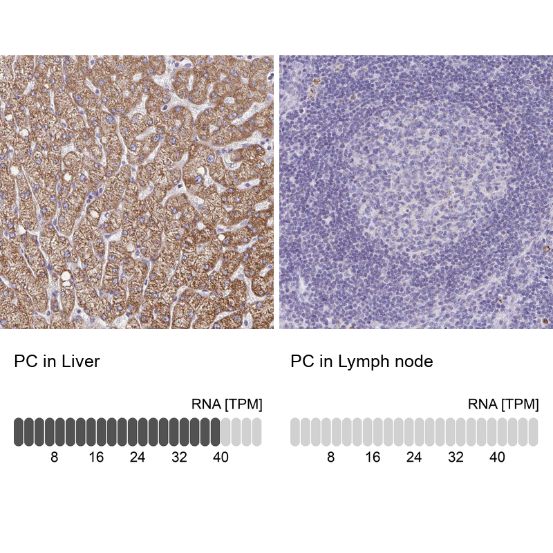
- Immunohistochemistry analysis in human liver and lymph node tissues using HPA043922 antibody. Corresponding PC RNA-seq data are presented for the same tissues.
- Sample type
- Human
- Protocol
- Protocol