Explore
 Validate
 Learn
Learn Western blot
Antibody data
- Antibody Data
- Antigen structure
- References [3]
- Comments [0]
- Validations
- Western blot [1]
- Immunohistochemistry [1]
Submit
Validation data
Reference
Comment
Report error
- Product number
- PAB11268 - Provider product page

- Provider
- Abnova Corporation
- Proper citation
- Abnova Corporation Cat#PAB11268, RRID:AB_1714586
- Product name
- SPANXC polyclonal antibody
- Antibody type
- Polyclonal
- Description
- Rabbit polyclonal antibody raised against full length recombinant SPANXC.
- Storage
- Store at 4°C. For long term storage store at -20°C.Aliquot after reconstitution to avoid repeated freezing and thawing.
Submitted references Dynamic structure of the SPANX gene cluster mapped to the prostate cancer susceptibility locus HPCX at Xq27.
The SPANX gene family of cancer/testis-specific antigens: rapid evolution and amplification in African great apes and hominids.
Genomic organization, incidence, and localization of the SPAN-x family of cancer-testis antigens in melanoma tumors and cell lines.
Kouprina N, Pavlicek A, Noskov VN, Solomon G, Otstot J, Isaacs W, Carpten JD, Trent JM, Schleutker J, Barrett JC, Jurka J, Larionov V
Genome research 2005 Nov;15(11):1477-86
Genome research 2005 Nov;15(11):1477-86
The SPANX gene family of cancer/testis-specific antigens: rapid evolution and amplification in African great apes and hominids.
Kouprina N, Mullokandov M, Rogozin IB, Collins NK, Solomon G, Otstot J, Risinger JI, Koonin EV, Barrett JC, Larionov V
Proceedings of the National Academy of Sciences of the United States of America 2004 Mar 2;101(9):3077-82
Proceedings of the National Academy of Sciences of the United States of America 2004 Mar 2;101(9):3077-82
Genomic organization, incidence, and localization of the SPAN-x family of cancer-testis antigens in melanoma tumors and cell lines.
Westbrook VA, Schoppee PD, Diekman AB, Klotz KL, Allietta M, Hogan KT, Slingluff CL, Patterson JW, Frierson HF, Irvin WP Jr, Flickinger CJ, Coppola MA, Herr JC
Clinical cancer research : an official journal of the American Association for Cancer Research 2004 Jan 1;10(1 Pt 1):101-12
Clinical cancer research : an official journal of the American Association for Cancer Research 2004 Jan 1;10(1 Pt 1):101-12
No comments: Submit comment
Supportive validation
- Submitted by
- Abnova Corporation (provider)
- Main image
- Experimental details
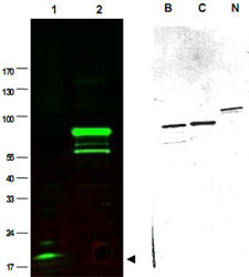
- Western blot using SPANXC polyclonal antibody (Cat # PAB11268) shows detection of a band at ~17 KDa corresponding to SPANXC present in a nuclear extract from VWM105 cells (left panel, arrowhead).VWM105 cells are derived from a human melanoma and are positive for SPANX proteins.Lane 2 shows reactivity with a purified recombinant SPANXC fusion protein.The right panel shows similar reactivity with purified recombinant SPANXB, SPANXC and SPANXN proteins.Proteins were separated by SDS-PAGE, transferred to nitrocellulose, and probed with the primary antibody diluted to 1 : 1,000.IRDye™800 conjugated Gt-a-Rabbit IgG [H&L] MX was used (left).IRDye is a trademark of LI-COR, Inc.Size estimation was made by comparison to prestained MW markers as indicated.Personal Communication. Vladimir Larionov, NIH, CCR, Bethesda, MD.

Supportive validation
- Submitted by
- Abnova Corporation (provider)
- Main image
- Experimental details
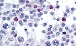
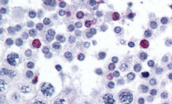
- Immunohistochemistry of SPANXC polyclonal antibody (Cat # PAB11268) was used at 2.5 ug/mL to detect signal in a variety of tissues including multi-human, multi-brain and multi-cancer slides.This image shows moderate positive staining of human sperm and spermatids at 60X.Tissue was formalin-fixed and paraffin embedded.The image shows localization of the antibody as the precipitated red signal, with a hematoxylin purple nuclear counterstain.Personal Communication, Tina Roush, Life Span Biosciences, Seattle, WA.
- Validation comment
- Immunohistochemistry (Formalin/PFA-fixed paraffin-embedded sections)