 Explore
 Validate
 Learn
Learn Flow cytometry
Antibody data
- Antibody Data
- Antigen structure
- References [6]
- Comments [0]
- Validations
- Flow cytometry [1]
- Other assay [8]
Submit
Validation data
Reference
Comment
Report error
- Product number
- 17-9117-42 - Provider product page

- Provider
- Invitrogen Antibodies
- Product name
- CD268 (BAFF Receptor) Monoclonal Antibody (8A7), APC, eBioscience™
- Antibody type
- Monoclonal
- Antigen
- Other
- Description
- Description: The 8A7 monoclonal antibody reacts with the human BAFF receptor, also known as B lymphocyte stimulator (BLyS protein) receptor, which is expressed on B cells. The ligand for this receptor, BAFF (B cell-activating factor of the TNF family) is a B cell survival factor and regulates CD21/35 and CD23 expression. Interaction of this ligand with its receptor causes elevated CD21/35 and CD23 expression, whereas receptor blockade has been shown to down-modulate expression. Applications Reported: This 8A7 antibody has been reported for use in flow cytometric analysis. Applications Tested: This 8A7 antibody has been pre-titrated and tested by flow cytometric analysis of normal human peripheral blood cells. This can be used at 5 µL (0.015 µg) per test. A test is defined as the amount (µg) of antibody that will stain a cell sample in a final volume of 100 µL. Cell number should be determined empirically but can range from 10^5 to 10^8 cells/test. Excitation: 633-647 nm; Emission: 660 nm; Laser: Red Laser. Filtration: 0.2 µm post-manufacturing filtered.
- Reactivity
- Human
- Host
- Mouse
- Isotype
- IgG
- Antibody clone number
- 8A7
- Vial size
- 100 Tests
- Concentration
- 5 µL/Test
- Storage
- 4° C, store in dark, DO NOT FREEZE!
Submitted references Altered expression profile of BAFF receptors on peripheral blood B lymphocytes in Graves' disease.
Type I Interferon Potentiates IgA Immunity to Respiratory Syncytial Virus Infection During Infancy.
IFN type I and II induce BAFF secretion from human decidual stromal cells.
BAFF promotes proliferation of human mesangial cells through interaction with BAFF-R.
The strength of the antibody response to the nematode Ascaris lumbricoides inversely correlates with levels of B-Cell Activating Factor (BAFF).
A functional receptor for B-cell-activating factor is expressed on human acute lymphoblastic leukemias.
Wang X, Huang J, Zhang A, Fang C, Ma Q, Jiang P
BMC endocrine disorders 2021 Apr 29;21(1):88
BMC endocrine disorders 2021 Apr 29;21(1):88
Type I Interferon Potentiates IgA Immunity to Respiratory Syncytial Virus Infection During Infancy.
Hijano DR, Siefker DT, Shrestha B, Jaligama S, Vu LD, Tillman H, Finkelstein D, Saravia J, You D, Cormier SA
Scientific reports 2018 Jul 23;8(1):11034
Scientific reports 2018 Jul 23;8(1):11034
IFN type I and II induce BAFF secretion from human decidual stromal cells.
Lundell AC, Nordström I, Andersson K, Lundqvist C, Telemo E, Nava S, Kaipe H, Rudin A
Scientific reports 2017 Jan 6;7:39904
Scientific reports 2017 Jan 6;7:39904
BAFF promotes proliferation of human mesangial cells through interaction with BAFF-R.
Zheng N, Wang D, Ming H, Zhang H, Yu X
BMC nephrology 2015 May 15;16:72
BMC nephrology 2015 May 15;16:72
The strength of the antibody response to the nematode Ascaris lumbricoides inversely correlates with levels of B-Cell Activating Factor (BAFF).
Bornacelly A, Mercado D, Acevedo N, Caraballo L
BMC immunology 2014 Jun 7;15:22
BMC immunology 2014 Jun 7;15:22
A functional receptor for B-cell-activating factor is expressed on human acute lymphoblastic leukemias.
Parameswaran R, Müschen M, Kim YM, Groffen J, Heisterkamp N
Cancer research 2010 Jun 1;70(11):4346-56
Cancer research 2010 Jun 1;70(11):4346-56
No comments: Submit comment
Supportive validation
- Submitted by
- Invitrogen Antibodies (provider)
- Main image

- Experimental details
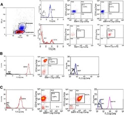
- Staining of normal human peripheral blood cells with Anti-Human CD19 eFluor® 450 (Product # 48-0199-42) and Mouse IgG2a K Isotype Control APC (Product # 17-4724-81) (left) or Anti-Human CD268 (BAFF Receptor) APC (right). Cells in the lymphocyte gate were used for analysis.

Supportive validation
- Submitted by
- Invitrogen Antibodies (provider)
- Main image
- Experimental details
- NULL
- Submitted by
- Invitrogen Antibodies (provider)
- Main image
- Experimental details
- NULL

- Submitted by
- Invitrogen Antibodies (provider)
- Main image
- Experimental details
- NULL

- Submitted by
- Invitrogen Antibodies (provider)
- Main image
- Experimental details
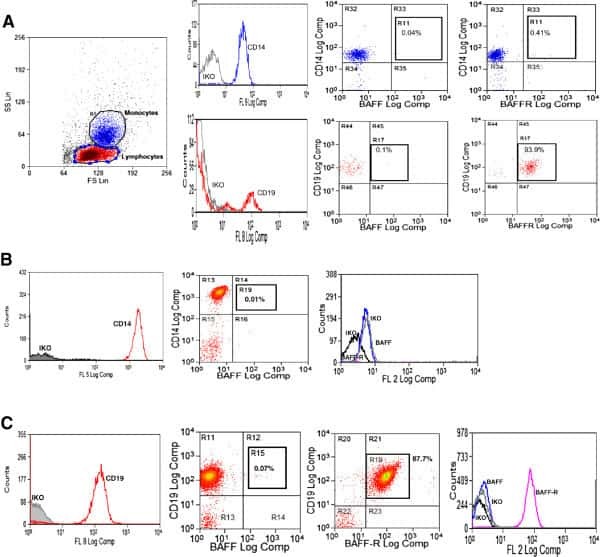
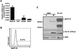
- Fig. 3 The expression of BAFF receptors by human mesangial cells. a Total RNA was extracted from human mesangial cells and analyzed for expression of BAFF-R, TACI, BCMA and BAFF, normalized to GAPDH expression, by real-time PCR. b The presence of BAFF-R on the surface of human mesangial cells was detected with BAFF-R antibody by flow cytometry. Background signal is presented as a gray line, and BAFF-R signal is presented as a blue line. c Cytosolic protein fraction (indicated as c) and membrane protein fraction (indicated as m) were prepared from human mesangial cells and subjected to western blot analysis. Whole protein lysate of activated human CD19 + B cells was loaded as a positive control for expression of BAFF-R. Na + /K + -ATPase and Erk kinase represented membrane and cytosolic proteins respectively
- Submitted by
- Invitrogen Antibodies (provider)
- Main image

- Experimental details
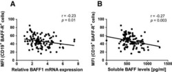
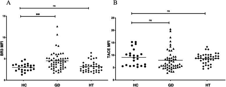
- Fig. 2 Altered expression profile of receptors of BAFF on peripheral blood B lymphocytes in GD. a : Elevated BR3 expression on peripheral blood B lymphocytes in GD patients. Mean MFI of BR3 on peripheral B lymphocytes in GD patients was 4.52 +- 2.06, higher than that in HC (3.00 +- 0.87), P = 0.0015; BR3 expression in HT patients was not increased; b : Trend of decreased TACI expression on peripheral B lymphocytes in GD patients. Mean MFI of TACI on peripheral B lymphocytes in GD patients was 7.96 +- 4.06, lower than that in HC (9.10 +- 3.37) without significance, P = 0.1285. TACI expression in HT was not decreased. MFI: mean fluorescence intensity. Bars: mean MFI. ns: not significant; **: vs. HC, P < 0.01
- Submitted by
- Invitrogen Antibodies (provider)
- Main image
- Experimental details
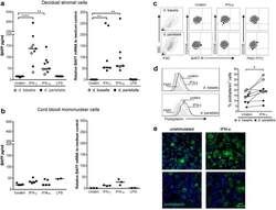
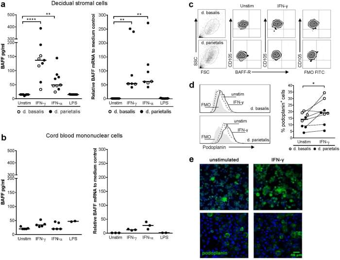
- Figure 3 IFN-gamma and IFN-alpha trigger BAFF production from decidual stromal cells. (a) BAFF production and relative BAFF mRNA levels to medium control by isolated decidual stromal cells in response to IFN-gamma, IFN-alpha or LPS. (b) BAFF production and relative BAFF mRNA levels to medium control by cord blood mononuclear cells in response to IFN-gamma, IFN-alpha or LPS. (c) BAFF-R expression on CD105-positive stromal cells isolated from decidua basalis or decidua parietalis after culture in the presence or absence of IFN-gamma. (d) Histograms depict podoplanin expression on stromal cells isolated from decidua basalis or decidua parietalis and the scatter plot shows the percentage of podoplanin-positive cells after stimulation with IFN-gamma or not. (e) Immunofluorescence staining of podoplanin on stromal cells isolated from decidua basalis stimulated with IFN-gamma or not (podoplanin in green and nuclei staining with Hoechst in blue). Images from one donor in the upper panel are presented with x 10 magnification and images from a second donor in the lower panel are presented with x 25 magnification (scale bar 40 mum). Horizontal bars indicate median. * P
- Submitted by
- Invitrogen Antibodies (provider)
- Main image

- Experimental details
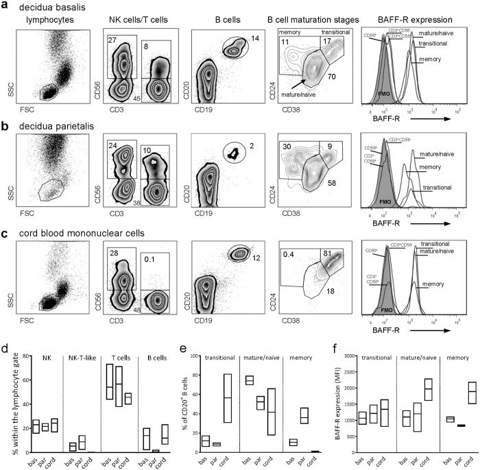
- Figure 4 BAFF-R expressing B cells at different maturational stages are present in decidua. To identify lymphocytes, in the first panel to the far left, live cells were gated using a viability dye that marks all dead cells and the lymphocyte gate was then set within CD45-expressing singlet leukocytes. (a - c) Within lymphocytes, CD56 + NK cells, CD3 + CD56 neg T cells, CD3 + CD56 + NK-T-like cells and CD19 + CD20 + B cells were identified (second and third panels). B cell maturational stages were distinguished based on CD24 and CD38 expression (fourth panel). The BAFF-R expression on the different lymphocyte populations is depicted to the far right. (d) The proportion of different cell subsets with in the lymphocyte gate and (e) the proportion of B cells at different maturational stages isolated from decidua basalis, decidua parietalis and cord blood. (f) The mean fluorescence intensity of BAFF on B cells at different maturational stages. Floating bars show minimum to maximum values, and horizontal bar indicates the median (n = 3). Approximately 40.000 cells were collected in the lymphocyte gate for d. basalis and CBMC and 20.000 for d. parietalis.
- Submitted by
- Invitrogen Antibodies (provider)
- Main image
- Experimental details
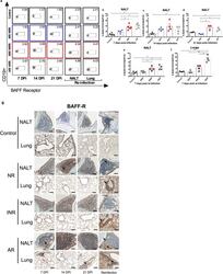
- Figure 4 BAFF-R-positive cells are increased in NALT by IFN-alpha administration or age in NALT upon primary infection with RSV. BAFF-R-positive cells by flow cytometry in NALT and lungs from RSV-infected adult (AR) and neonatal mice receiving IFN-alpha (INR) or placebo (NR) 16 h before RSV infection and adult (ARR) and neonatal mice receiving IFN-alpha (INRR) or placebo (NRR) 16 h before RSV infection and reinfected 4 weeks after primary infection. ( a ) Representative flow cytometry panels of B220 + CD19 + BAFF-R + cells. ( b-f ) Percentage of B220 + CD19 + BAFF-R + cells at different time points. ( b ) 7 dpi; ( c ) 14 dpi; ( d ) 21 dpi; ( e ) 7 days post-secondary infection in NALT; ( f ) 7 days post-secondary infection in lungs. ( g ) IHC analysis demonstrating expression of BAFF-R protein in NALT and lungs of RSV-challenged groups and control mice at 7, 14, and, 21 days post infection, as well as, after reinfection. Magnification 200x; scale bar = 100 mum. n = 3-5; N = 2 * P < 0.05, ** P < 0.01.
