Explore
 Validate
 Learn
Learn Western blot
 Immunocytochemistry
Antibody data
- Antibody Data
- Antigen structure
- References [1]
- Comments [0]
- Validations
- Immunocytochemistry [1]
- Other assay [2]
Submit
Validation data
Reference
Comment
Report error
- Product number
- PA5-106118 - Provider product page

- Provider
- Invitrogen Antibodies
- Product name
- Axl Polyclonal Antibody
- Antibody type
- Polyclonal
- Antigen
- Synthetic peptide
- Description
- Antibody detects endogenous levels of total AXL.
- Reactivity
- Human, Mouse, Rat
- Host
- Rabbit
- Isotype
- IgG
- Vial size
- 100 µL
- Concentration
- 1 mg/mL
- Storage
- -20°C
Submitted references TAM kinase signaling is indispensable for proper skeletal muscle regeneration in mice.
Al-Zaeed N, Budai Z, Szondy Z, Sarang Z
Cell death & disease 2021 Jun 12;12(6):611
Cell death & disease 2021 Jun 12;12(6):611
No comments: Submit comment
Supportive validation
- Submitted by
- Invitrogen Antibodies (provider)
- Main image

- Experimental details
- Immunofluorescent analysis of Axl in HUVEC cell lysate. Samples were fixed with paraformaldehyde, permeabilized with 0.1% Triton X-100, blocked with 10% serum (45 min at 25°C) incubated with Axl polyclonal antibody (Product # PA5-106118) using a dilution of 1:200 (1 hr, 37°C), and followed by goat anti-rabbit IgG Alexa Fluor 594 at a dilution of 1:600.

Supportive validation
- Submitted by
- Invitrogen Antibodies (provider)
- Main image

- Experimental details
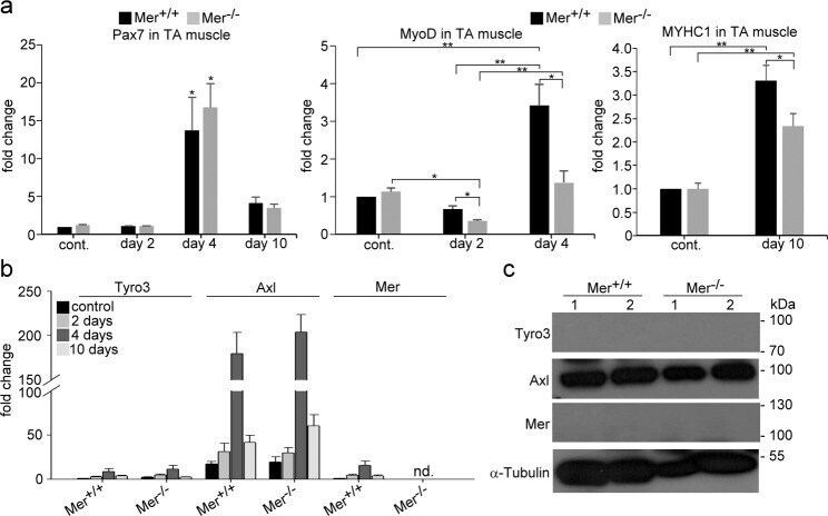
- Fig. 2 Expression of TAM kinase family members and myogenic genes in the TA muscle of wild type and Mer mice. a mRNA expressions of myogenic marker genes Pax7, MyoD and MYHC1 in control and in regenerating wild type and Mer TA muscles determined by qRT-PCR at day 2, 4, or 10 post-CTX-induced injury. b mRNA expression levels of Mer, Axl and Tyro3 in control and in regenerating wild type and Mer TA muscles determined by qRT-PCR at day 2, 4, or 10 post-CTX-induced injury. c Protein levels of Mer, Axl, and Tyro3 in the TA muscle of wild-type and Mer mice determined by Western blot analysis. alpha Tauubulin was used as a loading control. Data are expressed as mean +- SEM ( n = 3). Asterisks indicate statistical significance (* P < 0.05, ** P < 0.01, ANOVA test).
- Submitted by
- Invitrogen Antibodies (provider)
- Main image
- Experimental details
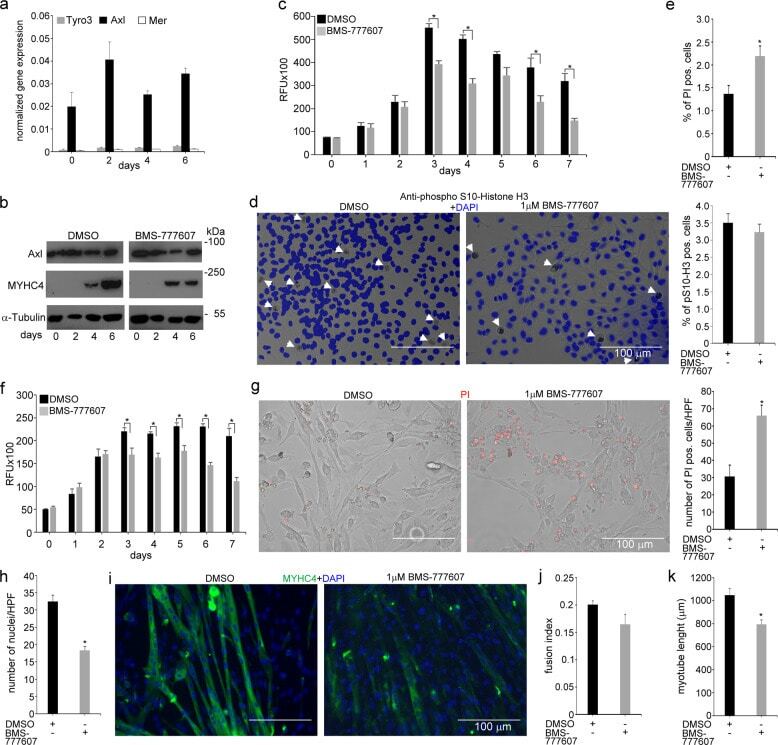
- Fig. 6 In vitro administration of the pan-TAM tyrosine kinase inhibitor BMS-777607 impairs myogenesis of C2C12 myoblast cells. a mRNA expression levels of Tyro3, Axl, and Mer during the differentiation of C2C12 myoblast cells determined by qRT-PCR. b Protein expression levels of Axl and myosin heavy chain 4 (MYHC4) in differentiating C2C12 myoblasts in the presence or absence of 1 muM BMS-777607 determined by Western blot analysis. alpha-Tubulin was used as a loading control. One representative blot of three is shown. c Alterations in the number of viable C2C12 cells grown in growth medium in the presence or absence of 1 muM BMS-777607 determined by PrestoBlue staining. d Percent of cells in G2/M phase grown in growth medium in the presence or absence of 1 muM BMS-777607 as indicated by anti-phospho-histone H3 (Ser10) and DAPI co-staining (at least 20 HPF were analyzed). Arrows point to the anti-phospho-histone H3 positive nuclei. e Percent of PI-positive cells grown in growth medium in the presence or absence of 1 muM BMS-777607. f Alterations in the number of viable C2C12 cells grown in differentiation medium in the presence or absence of 1 muM BMS-777607 determined by PrestoBlue staining. BMS-777607 was added when the medium was changed to differentiation medium. g Representative light microscopic images of C2C12 myoblasts differentiated for 6 days in the absence or presence of 1 muM BMS-777607 after staining dead cells with propidium iodide and quantification of propidium