 Explore
 Validate
 Learn
Learn Western blot
Antibody data
- Antibody Data
- Antigen structure
- References [12]
- Comments [0]
- Validations
- Western blot [1]
- Immunocytochemistry [1]
- Immunohistochemistry [1]
Submit
Validation data
Reference
Comment
Report error
- Product number
- AF2228 - Provider product page

- Provider
- R&D Systems
- Product name
- Human Phospho-Axl (Y779) Antibody
- Antibody type
- Polyclonal
- Description
- Antigen and protein A Affinity-purified. Detects human and mouse Axl when phosphorylated at Y779.
- Reactivity
- Human
- Host
- Rabbit
- Conjugate
- Unconjugated
- Isotype
- IgG
- Vial size
- 100 ug
- Concentration
- LYOPH
- Storage
- Use a manual defrost freezer and avoid repeated freeze-thaw cycles. 12 months from date of receipt, -20 to -70 °C as supplied. 1 month, 2 to 8 °C under sterile conditions after reconstitution. 6 months, -20 to -70 °C under sterile conditions after reconstitution.
Submitted references Induction of MET Receptor Tyrosine Kinase Down-regulation through Antibody-mediated Receptor Clustering.
AXL Mediates Esophageal Adenocarcinoma Cell Invasion through Regulation of Extracellular Acidification and Lysosome Trafficking.
DAPK and CIP2A are involved in GAS6/AXL-mediated Schwann cell proliferation in a rat model of bilateral cavernous nerve injury.
BET Inhibition Overcomes Receptor Tyrosine Kinase-Mediated Cetuximab Resistance in HNSCC.
Gas6 derived from cancer-associated fibroblasts promotes migration of Axl-expressing lung cancer cells during chemotherapy.
Tropomyosin isoform Tpm2.1 regulates collective and amoeboid cell migration and cell aggregation in breast epithelial cells.
Expression and role of TYRO3 and AXL as potential therapeutical targets in leiomyosarcoma.
Cell softening in malignant progression of human lung cancer cells by activation of receptor tyrosine kinase AXL.
Receptor tyrosine kinase Axl is required for resistance of leukemic cells to FLT3-targeted therapy in acute myeloid leukemia.
Identification of Axl as a downstream effector of TGF-β1 during Langerhans cell differentiation and epidermal homeostasis.
The novel receptor tyrosine kinase Axl is constitutively active in B-cell chronic lymphocytic leukemia and acts as a docking site of nonreceptor kinases: implications for therapy.
Growth arrest-specific protein 6 deficiency impairs liver tissue repair after acute toxic hepatitis in mice.
Li W, Dick A, Lu F, Zhang H, Sun H
Scientific reports 2019 Feb 13;9(1):1988
Scientific reports 2019 Feb 13;9(1):1988
AXL Mediates Esophageal Adenocarcinoma Cell Invasion through Regulation of Extracellular Acidification and Lysosome Trafficking.
Maacha S, Hong J, von Lersner A, Zijlstra A, Belkhiri A
Neoplasia (New York, N.Y.) 2018 Oct;20(10):1008-1022
Neoplasia (New York, N.Y.) 2018 Oct;20(10):1008-1022
DAPK and CIP2A are involved in GAS6/AXL-mediated Schwann cell proliferation in a rat model of bilateral cavernous nerve injury.
Chen YL, Tsai YT, Chao TT, Wu YN, Chen MC, Lin YH, Liao CH, Chou SP, Chiang HS
Oncotarget 2018 Jan 19;9(5):6402-6415
Oncotarget 2018 Jan 19;9(5):6402-6415
BET Inhibition Overcomes Receptor Tyrosine Kinase-Mediated Cetuximab Resistance in HNSCC.
Leonard B, Brand TM, O'Keefe RA, Lee ED, Zeng Y, Kemmer JD, Li H, Grandis JR, Bhola NE
Cancer research 2018 Aug 1;78(15):4331-4343
Cancer research 2018 Aug 1;78(15):4331-4343
Gas6 derived from cancer-associated fibroblasts promotes migration of Axl-expressing lung cancer cells during chemotherapy.
Kanzaki R, Naito H, Kise K, Takara K, Eino D, Minami M, Shintani Y, Funaki S, Kawamura T, Kimura T, Okumura M, Takakura N
Scientific reports 2017 Sep 6;7(1):10613
Scientific reports 2017 Sep 6;7(1):10613
Tropomyosin isoform Tpm2.1 regulates collective and amoeboid cell migration and cell aggregation in breast epithelial cells.
Shin H, Kim D, Helfman DM
Oncotarget 2017 Nov 10;8(56):95192-95205
Oncotarget 2017 Nov 10;8(56):95192-95205
Expression and role of TYRO3 and AXL as potential therapeutical targets in leiomyosarcoma.
Dantas-Barbosa C, Lesluyes T, Loarer FL, Chibon F, Treilleux I, Coindre JM, Meeus P, Brahmi M, Bally O, Ray-Coquard I, Sunyach MP, Cesne AL, Mir O, Bonvalot S, Toulmonde M, Italiano A, Saintigny P, Jean-Denis M, Ducimetiere F, Ranchere D, El Sayadi H, Alberti L, Blay JY
British journal of cancer 2017 Dec 5;117(12):1787-1797
British journal of cancer 2017 Dec 5;117(12):1787-1797
Cell softening in malignant progression of human lung cancer cells by activation of receptor tyrosine kinase AXL.
Iida K, Sakai R, Yokoyama S, Kobayashi N, Togo S, Yoshikawa HY, Rawangkan A, Namiki K, Suganuma M
Scientific reports 2017 Dec 19;7(1):17770
Scientific reports 2017 Dec 19;7(1):17770
Receptor tyrosine kinase Axl is required for resistance of leukemic cells to FLT3-targeted therapy in acute myeloid leukemia.
Park IK, Mundy-Bosse B, Whitman SP, Zhang X, Warner SL, Bearss DJ, Blum W, Marcucci G, Caligiuri MA
Leukemia 2015 Dec;29(12):2382-9
Leukemia 2015 Dec;29(12):2382-9
Identification of Axl as a downstream effector of TGF-β1 during Langerhans cell differentiation and epidermal homeostasis.
Bauer T, Zagórska A, Jurkin J, Yasmin N, Köffel R, Richter S, Gesslbauer B, Lemke G, Strobl H
The Journal of experimental medicine 2012 Oct 22;209(11):2033-47
The Journal of experimental medicine 2012 Oct 22;209(11):2033-47
The novel receptor tyrosine kinase Axl is constitutively active in B-cell chronic lymphocytic leukemia and acts as a docking site of nonreceptor kinases: implications for therapy.
Ghosh AK, Secreto C, Boysen J, Sassoon T, Shanafelt TD, Mukhopadhyay D, Kay NE
Blood 2011 Feb 10;117(6):1928-37
Blood 2011 Feb 10;117(6):1928-37
Growth arrest-specific protein 6 deficiency impairs liver tissue repair after acute toxic hepatitis in mice.
Lafdil F, Chobert MN, Deveaux V, Zafrani ES, Mavier P, Nakano T, Laperche Y, Brouillet A
Journal of hepatology 2009 Jul;51(1):55-66
Journal of hepatology 2009 Jul;51(1):55-66
No comments: Submit comment
Supportive validation
- Submitted by
- R&D Systems (provider)
- Main image
- Experimental details
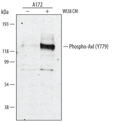
- Detection of Human Phospho-Axl (Y779) by Western Blot. Western blot shows lysates of A172 human glioblastoma cell line untreated (-) or treated (+) with WI38 human lung cell line conditioned media (WI38 CM) for 15 minutes. PVDF membrane was probed with 1 µg/mL of Human Phospho-Axl (Y779) Antigen Affinity-purified Polyclonal Antibody, followed by HRP-conjugated Anti-Rabbit IgG Secondary Antibody (Catalog # HAF008). A specific band was detected for Phospho-Axl (Y779) at approximately 140 kDa (as indicated). This experiment was conducted under reducing conditions and using Immunoblot Buffer Group 1.
Supportive validation
- Submitted by
- R&D Systems (provider)
- Main image
- Experimental details
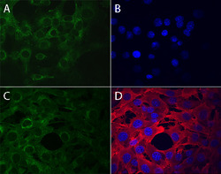
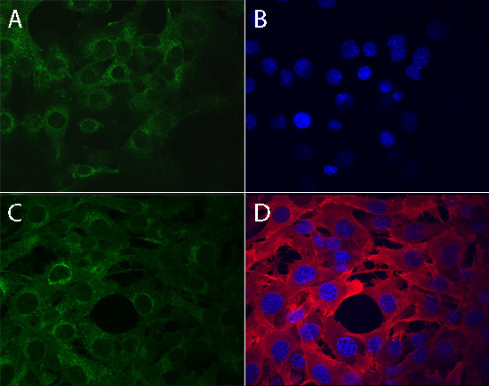
- Phospho-Axl (Y779) in A172 Human Cell Line. Axl phosphorylated at Y779 (panels B, D) and total Axl (panels A, C) were assessed in immersion fixed A172 human glioblastoma cells incubated with (panels C, D) or without (panels A, B) pervanadate. Phospho-Axl was detected using Rabbit Anti-Human Phospho-Axl (Y779) Antigen Affinity-purified Polyclonal Antibody (Catalog # AF2228) at 10 µg/mL for 3 hours at room temperature. Cells were stained using the NorthernLights™ 557-conjugated Anti-Rabbit IgG Secondary Antibody (red, panels B, D); Catalog # NL004) and counterstained using DAPI (blue). Total Axl was detected using Goat Anti-Human Axl Antigen Affinity-purified Polyclonal Antibody (Catalog # AF154). Cells were stained using the NorthernLights™ 493-conjugated Anti-Goat IgG Secondary Antibody (green, panels A, C); Catalog # NL003). Specific staining was localized to cytoplasm. View our protocol for Fluorescent ICC Staining of Cells on Coverslips.
Supportive validation
- Submitted by
- R&D Systems (provider)
- Main image
- Experimental details
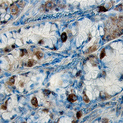
- Phospho-Axl (Y779) in Human Stomach Cancer Tissue. Axl phosphorylated at site Y779 was detected in immersion fixed paraffin-embedded sections of human stomach cancer tissue using Human Phospho-Axl (Y779) Antigen Affinity-purified Polyclonal Antibody (Catalog # AF2228) at 15 µg/mL overnight at 4 °C. Tissue was stained using the Anti-Rabbit HRP-DAB Cell & Tissue Staining Kit (brown; Catalog # CTS005) and counterstained with hematoxylin (blue). Specific labeling was localized to the cytoplasm of epithelial cells. View our protocol for Chromogenic IHC Staining of Paraffin-embedded Tissue Sections.
