 Explore
 Validate
 Learn
Learn Western blot
 Immunocytochemistry
Antibody data
- Antibody Data
- Antigen structure
- References [2]
- Comments [0]
- Validations
- Immunocytochemistry [3]
- Immunohistochemistry [1]
Submit
Validation data
Reference
Comment
Report error
- Product number
- PA5-48204 - Provider product page

- Provider
- Invitrogen Antibodies
- Product name
- DHCR7 Polyclonal Antibody
- Antibody type
- Polyclonal
- Antigen
- Synthetic peptide
- Reactivity
- Human, Mouse
- Host
- Rabbit
- Isotype
- IgG
- Vial size
- 400 μL
- Concentration
- 0.5 mg/mL
- Storage
- Store at 4°C short term. For long term storage, store at -20°C, avoiding freeze/thaw cycles.
Submitted references Sterol and oxysterol synthases near the ciliary base activate the Hedgehog pathway.
Endothelial cells control vascular smooth muscle cell cholesterol levels by regulating 24-dehydrocholesterol reductase expression.
Findakly S, Daggubati V, Garcia G, LaStella SA, Choudhury A, Tran C, Li A, Tong P, Garcia JQ, Puri N, Reiter JF, Xu L, Raleigh DR
The Journal of cell biology 2021 Jan 4;220(1)
The Journal of cell biology 2021 Jan 4;220(1)
Endothelial cells control vascular smooth muscle cell cholesterol levels by regulating 24-dehydrocholesterol reductase expression.
Kohlhaas J, Jäger MA, Lust L, De La Torre C, Hecker M, Korff T
Experimental cell research 2021 Feb 15;399(2):112446
Experimental cell research 2021 Feb 15;399(2):112446
No comments: Submit comment
Supportive validation
- Submitted by
- Invitrogen Antibodies (provider)
- Main image
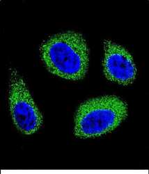
- Experimental details
- Immunocytochemistry analysis of DHCR7 in MCF-7 cells. Samples were incubated in DHCR7 polyclonal antibody (Product # PA5-48204) followed by Alexa Fluor 488-conjugated goat anti-rabbit lgG (green). DAPI was used to stain the cell nuclear (blue).

- Submitted by
- Invitrogen Antibodies (provider)
- Main image
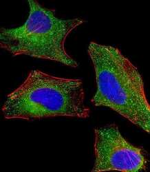
- Experimental details
- Immunofluorecent analysis of DHCR7 in HeLa cells. Cells were fixed, permeabilized and stained with DHCR7 polyclonal Antibody (Product # PA5-48204) at a 1:25 dilution, followed by Dylight® 488-conjugated goat anti-rabbit IgG secondary antibody at 1:200 dilution (green). Cytoplasmic actin is detected with Dylight® 554 Phalloidin at 1:100 dilution (red). The nuclear counter stain is DAPI (blue).

- Submitted by
- Invitrogen Antibodies (provider)
- Main image

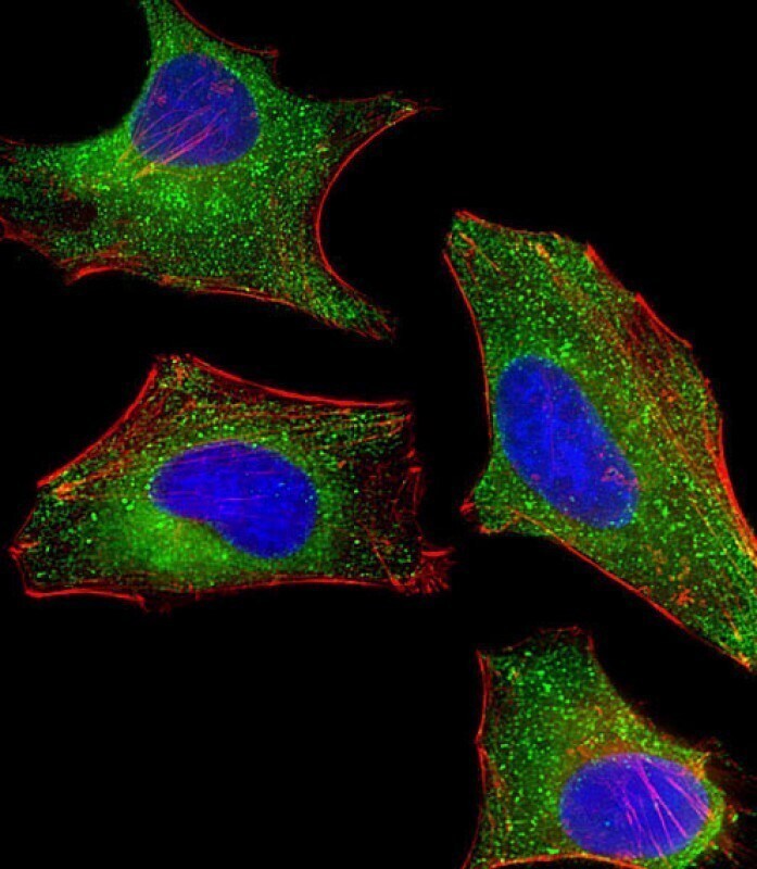
- Experimental details
- Immunocytochemistry analysis of DHCR7 in Hela (Human Cervical epithelial adenocarcinoma cell line) cells. Samples were incubated with DHCR7 polyclonal antibody (Product # PA5-48204) using a dilution of 1:25 followed by Dylight® 488-conjugated goat anti-rabbit IgG at a dilution of 1:200 (green). Cells were 4% paraformaldehyde-fixed, 0. 1% Triton X-100 permeabilized. Immunofluorescence image showing cytoplasm staining on HeLa cell line. Cytoplasmic actin is detected with Dylight® 554 Phalloidin at 1:100 dilution (red). The nuclear counter stain is DAPI (blue).
Supportive validation
- Submitted by
- Invitrogen Antibodies (provider)
- Main image
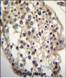
- Experimental details
- Immunohistochemistry analysis of DHCR7 in formalin fixed and paraffin embedded human testis tissue. Samples were incubated with DHCR7 polyclonal antibody (Product # PA5-48204) followed by peroxidase conjugation of the secondary antibody and DAB staining. This data demonstrates the use of this antibody for immunohistochemistry. Clinical relevance has not been evaluated.
