Explore
 Validate
 Learn
Learn Western blot
 Immunohistochemistry
Antibody data
- Antibody Data
- Antigen structure
- References [4]
- Comments [0]
- Validations
- Immunohistochemistry [1]
- Other assay [6]
Submit
Validation data
Reference
Comment
Report error
- Product number
- PA5-33875 - Provider product page

- Provider
- Invitrogen Antibodies
- Product name
- KCNN4 Polyclonal Antibody
- Antibody type
- Polyclonal
- Antigen
- Synthetic peptide
- Description
- Percent identity with other species by BLAST analysis: Human, Gorilla, Bovine (100%) Marmoset, Panda, Dog, Rabbit, Pig (93%) Mouse, Rat, Guinea pig (86%).
- Reactivity
- Human, Mouse, Bovine
- Host
- Rabbit
- Isotype
- IgG
- Vial size
- 50 μg
- Concentration
- 0.71 mg/mL
- Storage
- Store at 4°C short term. For long term storage, store at -20°C, avoiding freeze/thaw cycles.
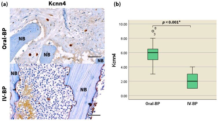
Submitted references Comparison of the Effect of Oral Versus Intravenous Bisphosphonate Administration on Osteoclastogenesis in Advanced-Stage Medication-Related Osteonecrosis of the Jaw Patients.
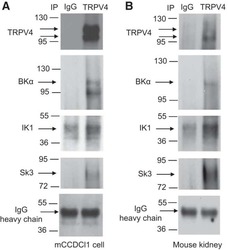
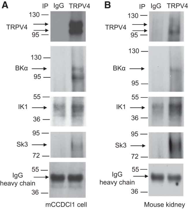
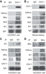
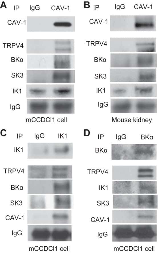
Caveolae facilitate TRPV4-mediated Ca(2+) signaling and the hierarchical activation of Ca(2+)-activated K(+) channels in K(+)-secreting renal collecting duct cells.
Expression of a Diverse Array of Ca2+-Activated K+ Channels (SK1/3, IK1, BK) that Functionally Couple to the Mechanosensitive TRPV4 Channel in the Collecting Duct System of Kidney.
Human neuronal changes in brain edema and increased intracranial pressure.
Kim HW, Lee MW, Lee JH, Kim MY
Journal of clinical medicine 2021 Jul 4;10(13)
Journal of clinical medicine 2021 Jul 4;10(13)
Caveolae facilitate TRPV4-mediated Ca(2+) signaling and the hierarchical activation of Ca(2+)-activated K(+) channels in K(+)-secreting renal collecting duct cells.
Li Y, Hu H, O'Neil RG
American journal of physiology. Renal physiology 2018 Dec 1;315(6):F1626-F1636
American journal of physiology. Renal physiology 2018 Dec 1;315(6):F1626-F1636
Expression of a Diverse Array of Ca2+-Activated K+ Channels (SK1/3, IK1, BK) that Functionally Couple to the Mechanosensitive TRPV4 Channel in the Collecting Duct System of Kidney.
Li Y, Hu H, Butterworth MB, Tian JB, Zhu MX, O'Neil RG
PloS one 2016;11(5):e0155006
PloS one 2016;11(5):e0155006
Human neuronal changes in brain edema and increased intracranial pressure.
Faragó N, Kocsis ÁK, Braskó C, Lovas S, Rózsa M, Baka J, Kovács B, Mikite K, Szemenyei V, Molnár G, Ozsvár A, Oláh G, Piszár I, Zvara Á, Patócs A, Barzó P, Puskás LG, Tamás G
Acta neuropathologica communications 2016 Aug 4;4(1):78
Acta neuropathologica communications 2016 Aug 4;4(1):78
No comments: Submit comment
Supportive validation
- Submitted by
- Invitrogen Antibodies (provider)
- Main image
- Experimental details
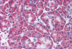
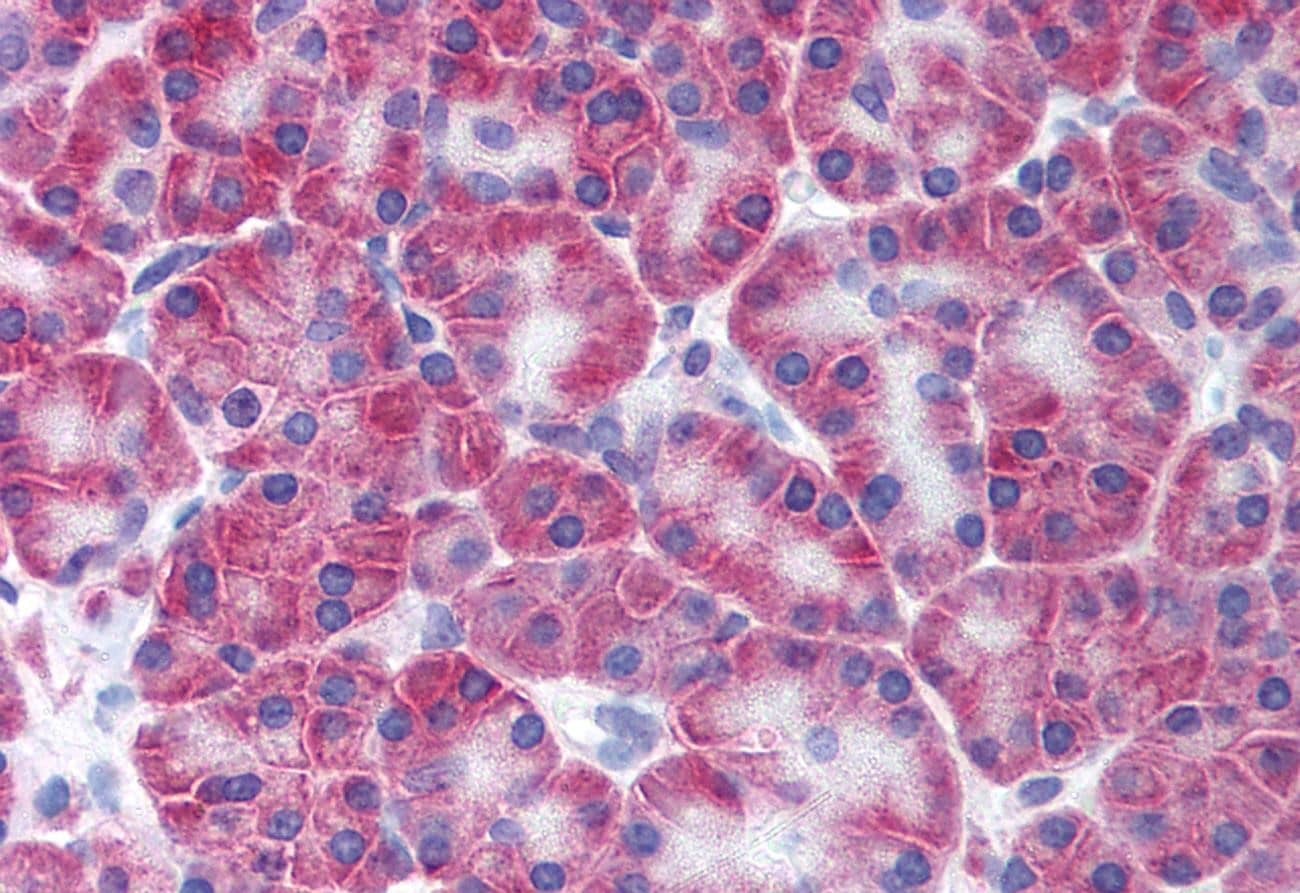
- Immunohistochemistry analysis of KCNN4 in human pancreas. Samples were incubated with KCNN4 polyclonal antibody (Product # PA5-33875). Formalin-fixed, paraffin-embedded tissue after heat-induced antigen retrieval.
Supportive validation
- Submitted by
- Invitrogen Antibodies (provider)
- Main image
- Experimental details
- NULL
- Submitted by
- Invitrogen Antibodies (provider)
- Main image
- Experimental details
- NULL
- Submitted by
- Invitrogen Antibodies (provider)
- Main image
- Experimental details
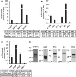
- Fig 1 Differential mRNA and protein expression levels of ion channels in mCCDcl1 cells. A. qPCR analysis of mRNA expression levels of marker channels relative to the AQP2 water channel. The mechanosensitive TRPV4 channel mRNA levels and ROMK mRNA levels are relatively high while a second mechanosensitive TRP channel, TRPV2, are very low. For comparison, mRNA expression levels for all channels are also given, relative to AQP2 mRNA levels, in the accompanying Table 1 . B. qPCR mRNA expression levels of KCa channels relative to the ROMK channel. It shows that SK1, SK3, IK1 are expressed at relative high mRNA levels, BKalpha at moderate mRNA levels, and SK2 at low mRNA levels. C. mRNA expression of BK channel subunits relative to BKalpha mRNA levels. It shows high relative mRNA levels for BKbeta1 and BKbeta2, but relatively low levels for BKbeta4. D. Immunoblots of mCCDcl1 cells for the key channels showing appropriate protein bands for TRPV4 (98 kD), SK1 (64 kD), SK3 (81 kD), IK1 (45 kD), and BKalpha (110 kD).

- Submitted by
- Invitrogen Antibodies (provider)
- Main image
- Experimental details
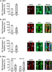
- Fig 6 Differential expression of ion channels between principal cells (PC) and intercalated cells (IC) in the mouse kidney CCD. AQP2 staining was used to identify PC, the AQP2-positive cells, and IC, the APQ2-negative cells. Bar graphs give the mean +- SEM for the normalized intensities for each KCa channel for both PC and IC. The number of PC and IC cells analyzed, n, is indicated on the bar graphs. The images of CCD show representative immunofluorescence examples for each KCa channel and AQP2 (40X) where one or two PC (AQP2-positive) and IC (AQP2-negative) cells are labelled as ""PC"" or ""IC."" The outer border of the tubule in each image is indicated by the dashed white line (indicating the basal side or anti-luminal side of tubular cells). ""L"" identifies the tubular lumen. The immunofluorescence intensity was determined for each KCa channel using ImageJ and normalized to the intensity levels of AQP2 expression in PC (see Methods ). Each example gives staining for AQP2 (red), the KCa channel (green), and the merged image (AQP2, KCa channel) that includes DAPI staining to identify nuclei. A. Expression of SK1 in PC and IC showing dominant relative expression in IC over PC (P
- Submitted by
- Invitrogen Antibodies (provider)
- Main image
- Experimental details
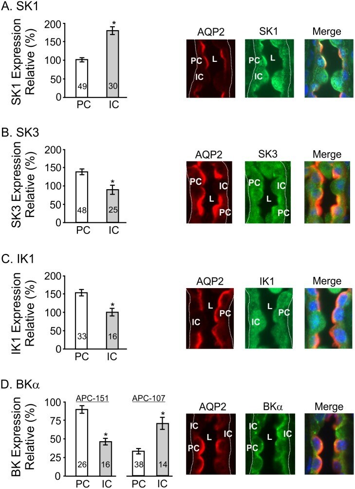
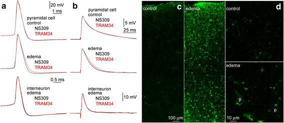
- Fig. 3 Functional validation of KCNN4 mRNA copy number changes in detected human pyramidal cells. Waveforms of action potentials evoked by depolarizing current injections ( a ) and of extracellularly evoked EPSPs ( b ) responded differently to the serial application of the small- and intermediate-conductance calcium activated potassium channel activator NS309 (500 nM) and TRAM34 (1 muM), an inhibitor of intermediate-conductance calcium activated potassium channels. The descending phase of action potentials and EPSPs was shortened in pyramidal cells recorded in brain slices prepared from the Edema group, but remained unchanged in pyramidal neurons of the Control group and in fast spiking interneurons of the Edema group. Traces shown are population averages. Confocal images of immunoreactions with antibodies against Kcnn4 performed simultaneously on samples of the Control and Edema groups showing a cross section of the gray matter ( c ) and part of layer 3 similar to areas where electrophysiological experiments were performed ( d ). Pyramidal cells were not labeled in the Control group and moderate Kcnn4 positivity was detected in pyramidal cells (p) of the Edema group. In addition, intense immunolabeling for Kcnn4 was detected in glial cells resembling astrocytes and interlaminar glia in both groups of patients
- Submitted by
- Invitrogen Antibodies (provider)
- Main image

- Experimental details
- Figure 3 Kcnn4 expression is decreased in the necrotic bones of IV BP-induced advanced-stage MRONJ patients. ( a ) High-magnification images (400x) of the immunohistochemistry analysis of Kcnn4 expression in the specimens of the two patient groups as indicated. Scale bars = 50 mum. Statistical analysis of the number of Kcnn4-positive cells ( b ), via the Mann-Whitney U test ( n = 20 per group). deg marks statistical outliers. For detailed data, see Table 2 . * p < 0.001.