Explore
 Validate
 Learn
Learn Western blot
Antibody data
- Antibody Data
- Antigen structure
- References [0]
- Comments [0]
- Validations
- Western blot [2]
- Immunocytochemistry [3]
- Immunohistochemistry [6]
- Flow cytometry [1]
Submit
Validation data
Reference
Comment
Report error
- Product number
- MA5-32914 - Provider product page

- Provider
- Invitrogen Antibodies
- Product name
- ALDH2 Monoclonal Antibody (E4-D10)
- Antibody type
- Monoclonal
- Antigen
- Synthetic peptide
- Reactivity
- Human, Mouse, Rat
- Host
- Mouse
- Isotype
- IgG
- Antibody clone number
- E4-D10
- Vial size
- 100 µL
- Concentration
- 2 mg/mL
- Storage
- Store at 4°C short term. For long term storage, store at -20°C, avoiding freeze/thaw cycles.
No comments: Submit comment
Supportive validation
- Submitted by
- Invitrogen Antibodies (provider)
- Main image

- Experimental details
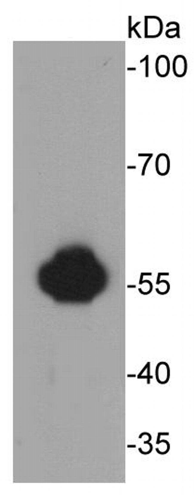
- Western blot analysis of ALDH2 in HepG2 cell lysates using a ALDH2 Monoclonal antibody (Product # MA5-32914).
- Submitted by
- Invitrogen Antibodies (provider)
- Main image

- Experimental details
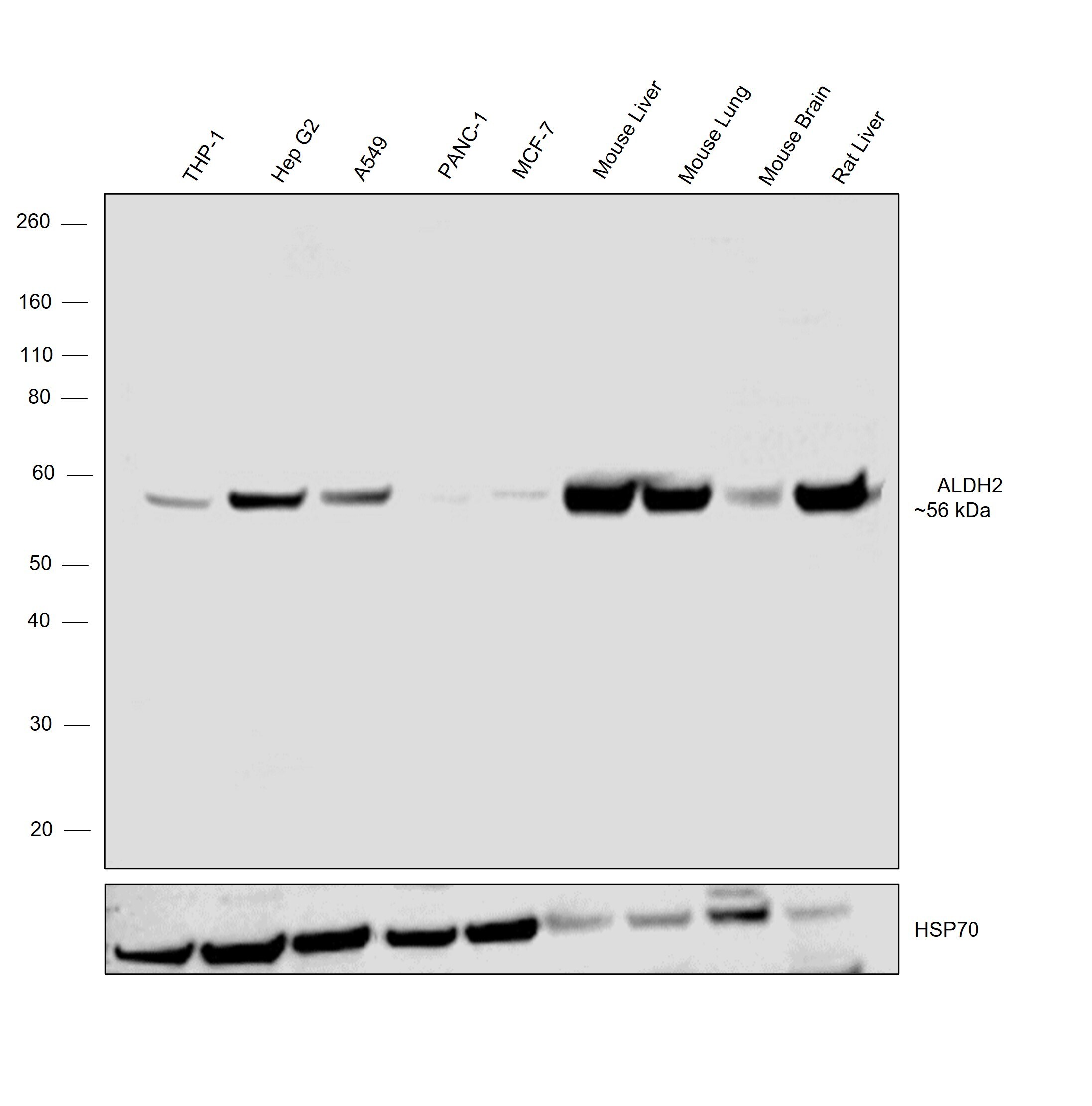
- Western blot was performed using Anti-ALDH2 Monoclonal Antibody (E4-D10) (Product # MA5-32914) and a 56 kDa band corresponding to acetaldehyde dehydrogenase 2; ALDH2 was observed across the panel tested except for PANC-1, MCF7 and mouse brain which are reported to be low expressors of ALDH2. Whole cell extracts (30 µg lysate) of THP-1 (Lane 1), Hep G2 (Lane 2), A549 (Lane 3), PANC-1 (Lane 4), MCF7 (Lane 5), Mouse Liver (Lane 6), Mouse Lung (Lane 7), Mouse Brain (Lane 8), Rat Liver (Lane 9) were electrophoresed using NuPAGE™ 4-12% Bis-Tris Protein Gel (Product # NP0322BOX), 12 well. Resolved proteins were then transferred onto a nitrocellulose membrane (Product # IB23001) by iBlot® 2 Dry Blotting System (Product # IB21001). The blot was probed with the primary antibody (1:1000 dilution) and detected by chemiluminescence with Goat anti-Mouse IgG (H+L) Superclonal™ Recombinant Secondary Antibody, HRP (Product # A28177, 1:20,000 dilution) using the iBright™ FL1500 Imaging System (Product # A44115). Chemiluminescent detection was performed using SuperSignal™ West Pico PLUS Chemiluminescent Substrate (Product # 34580).
Supportive validation
- Submitted by
- Invitrogen Antibodies (provider)
- Main image

- Experimental details
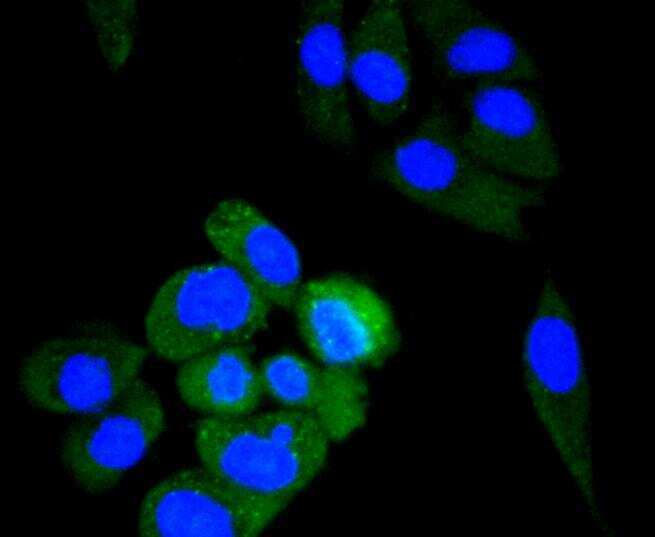
- Immunocytochemical analysis of ALDH2 in HepG2 cells using a ALDH2 Monoclonal antibody (Product # MA5-32914)..
- Submitted by
- Invitrogen Antibodies (provider)
- Main image

- Experimental details
- Immunocytochemical analysis of ALDH2 in PC-3M cells using a ALDH2 Monoclonal antibody (Product # MA5-32914)..
- Submitted by
- Invitrogen Antibodies (provider)
- Main image
- Experimental details
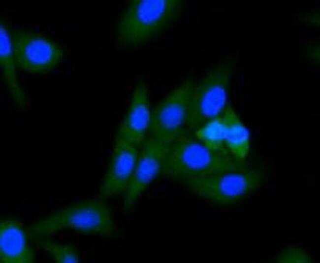
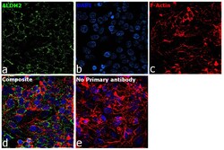
- Immunofluorescence analysis of acetaldehyde dehydrogenase 2; ALDH2 was performed using 70% confluent log phase Hep G2 cells. The cells were fixed with 4% paraformaldehyde for 10 minutes, permeabilized with 0.1% Triton™ X-100 for 15 minutes, and blocked with 2% BSA for 45 minutes at room temperature. The cells were labeled with ALDH2 Monoclonal Antibody (E4-D10) (Product # MA5-32914) at 1:200 dilution in 0.1% BSA, incubated at 4 degree celsius overnight and then labeled with Donkey anti-Rabbit IgG (H+L) Highly Cross-Adsorbed Secondary Antibody, Alexa Fluor Plus 488 (Product # A32790, 1:3000 dilution), for 45 minutes at room temperature (Panel a: Green). Nuclei (Panel b: Blue) were stained with ProLong™ Diamond Antifade Mountant with DAPI (Product # P36962). F-actin (Panel c: Red) was stained with Rhodamine Phalloidin (Product # R415, 1:300). Panel d represents the merged image showing mitochondria localization. Panel e represents control cells with no primary antibody to assess background. The images were captured at 60X magnification.

Supportive validation
- Submitted by
- Invitrogen Antibodies (provider)
- Main image

- Experimental details
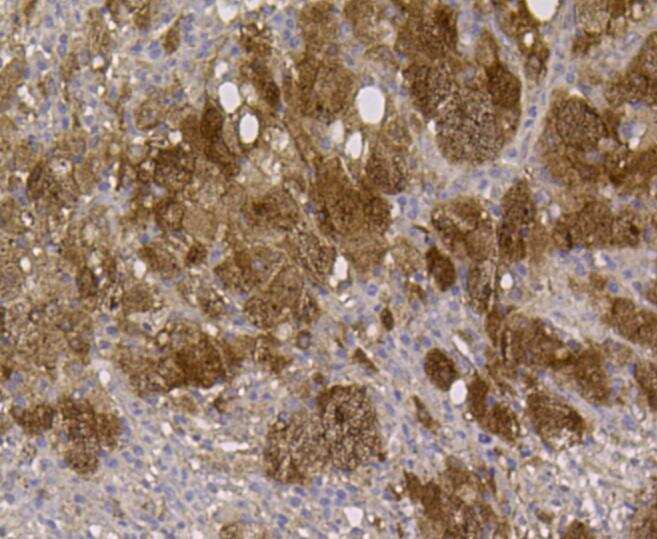
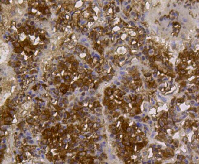
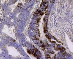
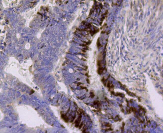
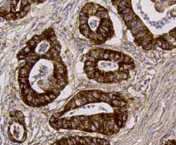
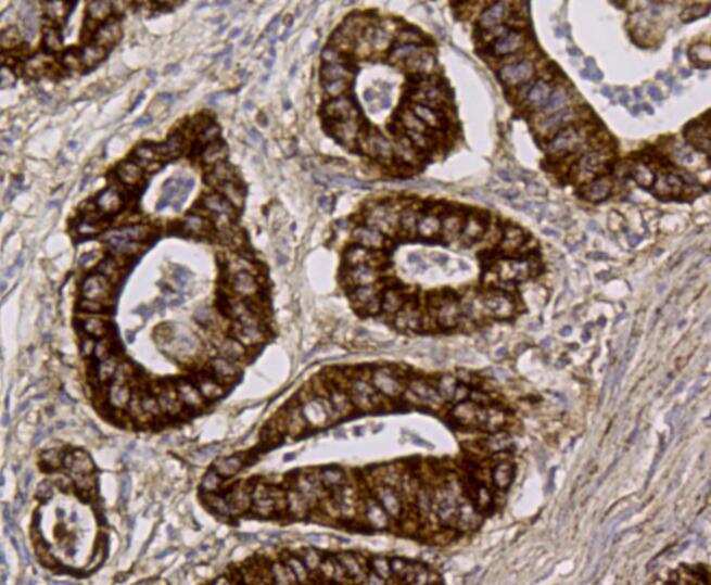
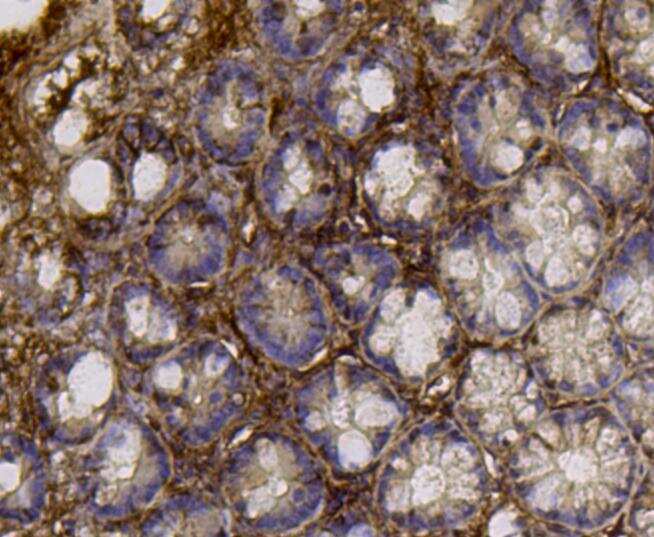
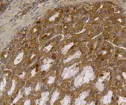
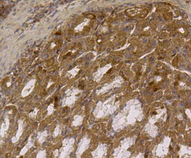
- Immunohistochemical analysis of ALDH2 of paraffin-embedded Human lung cancer tissue using a ALDH2 Monoclonal antibody (Product #MA5-32914).
- Submitted by
- Invitrogen Antibodies (provider)
- Main image
- Experimental details
- Immunohistochemical analysis of ALDH2 of paraffin-embedded Human lung cancer tissue using a ALDH2 Monoclonal antibody (Product #MA5-32914).
- Submitted by
- Invitrogen Antibodies (provider)
- Main image
- Experimental details
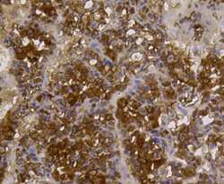
- Immunohistochemical analysis of ALDH2 of paraffin-embedded Human lung cancer tissue using a ALDH2 Monoclonal antibody (Product #MA5-32914).
- Submitted by
- Invitrogen Antibodies (provider)
- Main image
- Experimental details
- Immunohistochemical analysis of ALDH2 of paraffin-embedded Human lung cancer tissue using a ALDH2 Monoclonal antibody (Product #MA5-32914).
- Submitted by
- Invitrogen Antibodies (provider)
- Main image

- Experimental details
- Immunohistochemical analysis of ALDH2 of paraffin-embedded Human lung cancer tissue using a ALDH2 Monoclonal antibody (Product #MA5-32914).
- Submitted by
- Invitrogen Antibodies (provider)
- Main image
- Experimental details
- Immunohistochemical analysis of ALDH2 of paraffin-embedded Human lung cancer tissue using a ALDH2 Monoclonal antibody (Product #MA5-32914).
Supportive validation
- Submitted by
- Invitrogen Antibodies (provider)
- Main image
- Experimental details
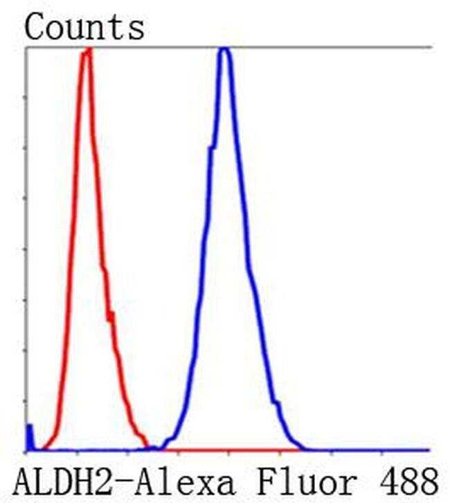
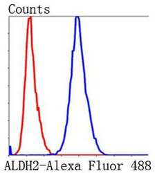
- Flow Cytometric analysis of ALDH2 in HepG2 cells using a ALDH2 Monoclonal Antibody (Product # MA5-32914) at a dilution of 1:100, as seen in blue compared with an unlabelled control (cells without incubation with primary antibody; red). Alexa Fluor 488-conjugated Goat anti mouse IgG was used as the secondary antibody.