Explore
 Validate
 Learn
Learn Western blot
 Immunocytochemistry
Antibody data
- Antibody Data
- Antigen structure
- References [3]
- Comments [0]
- Validations
- Immunocytochemistry [7]
- Immunohistochemistry [2]
- Other assay [2]
Submit
Validation data
Reference
Comment
Report error
- Product number
- PA5-21712 - Provider product page

- Provider
- Invitrogen Antibodies
- Product name
- WNT11 Polyclonal Antibody
- Antibody type
- Polyclonal
- Antigen
- Recombinant full-length protein
- Description
- Recommended positive controls: 293T, A431, HeLa, HepG2, mouse heart. Predicted reactivity: Mouse (96%), Rat (96%), Xenopus laevis (81%), Rabbit (97%), Chicken (85%), Bovine (98%). Store product as a concentrated solution. Centrifuge briefly prior to opening the vial.
- Reactivity
- Human, Mouse, Rat
- Host
- Rabbit
- Isotype
- IgG
- Vial size
- 100 μL
- Concentration
- 1.39 mg/mL
- Storage
- Store at 4°C short term. For long term storage, store at -20°C, avoiding freeze/thaw cycles.
Submitted references WNT11, a new gene associated with early onset osteoporosis, is required for osteoblastogenesis.
16p11.2 transcription factor MAZ is a dosage-sensitive regulator of genitourinary development.
Role of Noncanonical Wnt Signaling Pathway in Human Aortic Valve Calcification.
Caetano da Silva C, Edouard T, Fradin M, Aubert-Mucca M, Ricquebourg M, Raman R, Salles JP, Charon V, Guggenbuhl P, Muller M, Cohen-Solal M, Collet C
Human molecular genetics 2022 May 19;31(10):1622-1634
Human molecular genetics 2022 May 19;31(10):1622-1634
16p11.2 transcription factor MAZ is a dosage-sensitive regulator of genitourinary development.
Haller M, Au J, O'Neill M, Lamb DJ
Proceedings of the National Academy of Sciences of the United States of America 2018 Feb 20;115(8):E1849-E1858
Proceedings of the National Academy of Sciences of the United States of America 2018 Feb 20;115(8):E1849-E1858
Role of Noncanonical Wnt Signaling Pathway in Human Aortic Valve Calcification.
Albanese I, Yu B, Al-Kindi H, Barratt B, Ott L, Al-Refai M, de Varennes B, Shum-Tim D, Cerruti M, Gourgas O, Rhéaume E, Tardif JC, Schwertani A
Arteriosclerosis, thrombosis, and vascular biology 2017 Mar;37(3):543-552
Arteriosclerosis, thrombosis, and vascular biology 2017 Mar;37(3):543-552
No comments: Submit comment
Supportive validation
- Submitted by
- Invitrogen Antibodies (provider)
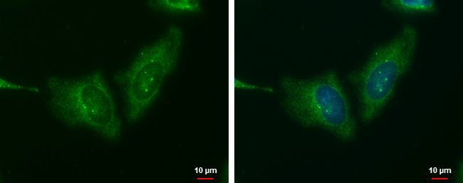
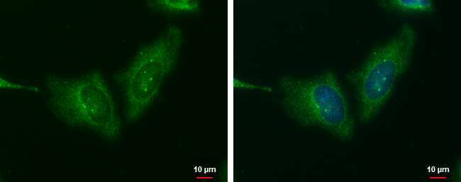
- Main image

- Experimental details
- Immunofluorescent analysis of WNT11 showing staining in the cytoplasm of HeLa cells. HeLa cells were fixed in ice-cold MeOH for 5 min and stained using a WNT11 polyclonal antibody (Product # PA5-21712) diluted at 1:500. Blue: Hoechst 33342 staining. Scale bar = 10µm.
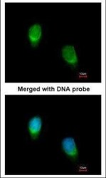
- Submitted by
- Invitrogen Antibodies (provider)
- Main image
- Experimental details
- Immunofluorescent analysis of WNT11 in paraformaldehyde-fixed HeLa cells using a WNT11 polyclonal antibody (Product # PA5-21712) at a 1:200 dilution.

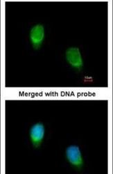
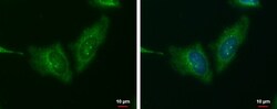
- Submitted by
- Invitrogen Antibodies (provider)
- Main image
- Experimental details
- WNT11 Polyclonal Antibody detects WNT11 protein at cytoplasm by immunofluorescent analysis. Sample: HeLa cells were fixed in ice-cold MeOH for 5 min. Green: WNT11 protein stained by WNT11 Polyclonal Antibody (Product # PA5-21712) diluted at 1:500. Blue: Hoechst 33342 staining. Scale bar = 10 µm.
- Submitted by
- Invitrogen Antibodies (provider)
- Main image
- Experimental details
- Immunofluorescence analysis of paraformaldehyde-fixed HeLa, using WNT11 antibody (Product # PA5-21712) at 1:200 dilution.

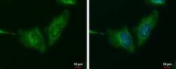
- Submitted by
- Invitrogen Antibodies (provider)
- Main image
- Experimental details
- WNT11 Polyclonal Antibody detects WNT11 protein at cytoplasm by immunofluorescent analysis. Sample: HeLa cells were fixed in ice-cold MeOH for 5 min. Green: WNT11 protein stained by WNT11 Polyclonal Antibody (Product # PA5-21712) diluted at 1:500. Blue: Hoechst 33342 staining. Scale bar = 10 µm.

- Submitted by
- Invitrogen Antibodies (provider)
- Main image

- Experimental details
- Immunofluorescence analysis of paraformaldehyde-fixed HeLa, using WNT11 antibody (Product # PA5-21712) at 1:200 dilution.

Supportive validation
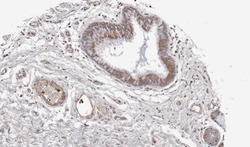
- Submitted by
- Invitrogen Antibodies (provider)
- Main image

- Experimental details
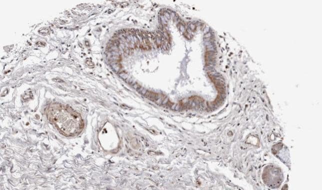
- WNT11 antibody detects WNT11 protein at secreted on mouse colon by immunohistochemical analysis. Sample: Paraffin-embedded mouse colon. WNT11 antibody (Product # PA5-21712) dilution: 1:500. Antigen Retrieval: EDTA based buffer, pH 8.0, 15 min.

- Submitted by
- Invitrogen Antibodies (provider)
- Main image
- Experimental details
- Immunohistochemical analysis of paraffin-embedded lung SCC xenograft, using WNT11 (Product # PA5-21712) antibody at 1:100 dilution. Antigen Retrieval: EDTA based buffer, pH 8.0, 15 min.
Supportive validation
- Submitted by
- Invitrogen Antibodies (provider)
- Main image
- Experimental details
- NULL
- Submitted by
- Invitrogen Antibodies (provider)
- Main image

- Experimental details
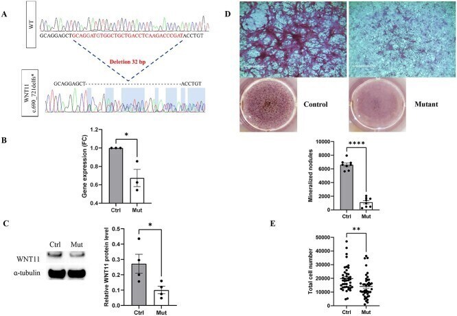
- Figure 2 WNT11 heterozygous mutant cells with decreased WNT11 mRNA and protein levels as well as proliferation and mineralization. ( A ) Electropherogram showing the 32-bp deletion leading to a frameshift. fs * : frameshift. ( B ) RT-qPCR analysis of WNT11 mRNA expression in mutant versus control U2OS cells. Normalization was to GAPDH level as a housekeeping gene with ratio of one for control U2OS cells. Ctrl: wild-type U2OS cells, Mut: WNT11 mutant cells, FC: fold change. ( C ) Western blot analysis of WNT11 protein expression, confirming the heterozygosity. a-Tubulin was a control. ( D ) Alizarin red staining showed formation of mineralized nodules after osteogenic differentiation treatment with osteogenic media in control and WNT11 mutant cells. ( E ) Proliferation of control and WNT11 mutant cells ( n = 42). Data are mean +- SEM. * P < 0.05, ** P < 0.01, **** P < 0.0001.