Explore
 Validate
 Learn
Learn Western blot
 ELISA
ELISAAntibody data
- Antibody Data
- Antigen structure
- References [1]
- Comments [0]
- Validations
- Western blot [3]
- Immunohistochemistry [1]
- Flow cytometry [1]
Submit
Validation data
Reference
Comment
Report error
- Product number
- NB100-1458 - Provider product page

- Provider
- Novus Biologicals
- Proper citation
- Novus Cat#NB100-1458, RRID:AB_2097337
- Product name
- Goat Polyclonal EHD2 Antibody
- Antibody type
- Polyclonal
- Description
- Immunogen affinity purified. This antibody is expected to recognise EHD1 protein as well as EHD2.
- Reactivity
- Human
- Host
- Goat
- Isotype
- IgG
- Vial size
- 0.1 mg
- Concentration
- 0.5 mg/ml
- Storage
- Store at -20C. Avoid freeze-thaw cycles.
Submitted references Role of EHD1 and EHBP1 in perinuclear sorting and insulin-regulated GLUT4 recycling in 3T3-L1 adipocytes.
Guilherme A, Soriano NA, Furcinitti PS, Czech MP
The Journal of biological chemistry 2004 Sep 17;279(38):40062-75
The Journal of biological chemistry 2004 Sep 17;279(38):40062-75
No comments: Submit comment
Supportive validation
- Submitted by
- Novus Biologicals (provider)
- Main image

- Experimental details
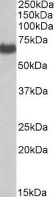
- Western Blot: EHD2 Antibody [NB100-1458] - Staining of Human Placenta lysate (35 ug protein in RIPA buffer). Primary incubation was 1 hour. Detected by chemiluminescence.
- Submitted by
- Novus Biologicals (provider)
- Main image

- Experimental details
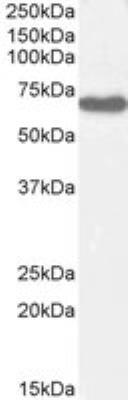
- Western Blot: EHD2 Antibody [NB100-1458] - Staining of Human Lung lysate with antibody at 0.3 ug/mL (35 ug protein in RIPA buffer). Detected by chemiluminescence.
- Submitted by
- Novus Biologicals (provider)
- Main image
- Experimental details
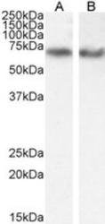
- Western Blot: EHD2 Antibody [NB100-1458] - Staining of A549 (A) with antibody at 0.1 ug/mL and HeLa (B) cell lysate with antibody at 0.3 ug/mL (35 ug protein in RIPA buffer). Detected by chemiluminescence.

Supportive validation
- Submitted by
- Novus Biologicals (provider)
- Main image
- Experimental details
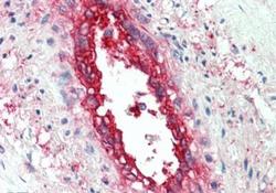
- Immunohistochemistry-Paraffin: EHD2 Antibody [NB100-1458] - Staining of paraffin embedded Human Vessel. Steamed antigen retrieval with citrate buffer pH 6, AP-staining.

Supportive validation
- Submitted by
- Novus Biologicals (provider)
- Main image
- Experimental details
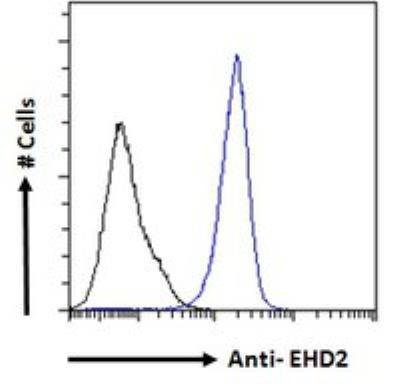
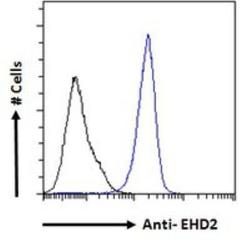
- Flow Cytometry: EHD2 Antibody [NB100-1458] - Flow cytometric analysis of paraformaldehyde fixed A431 cells (blue line), permeabilized with 0.5% Triton. Primary incubation 1hr (10 ug/mL) followed by Alexa Fluor 488 secondary antibody (1 ug/mL). IgG control: Unimmunized goat IgG (black line) followed by Alexa Fluor 488 secondary antibody.