 Explore
 Validate
 Learn
Learn Western blot
Antibody data
- Antibody Data
- Antigen structure
- References [1]
- Comments [0]
- Validations
- Western blot [1]
- Flow cytometry [1]
Submit
Validation data
Reference
Comment
Report error
- Product number
- MAB4379 - Provider product page

- Provider
- Novus Biologicals
- Product name
- Mouse Monoclonal TSC1 Antibody
- Antibody type
- Monoclonal
- Description
- Protein A or G purified from hybridoma culture supernatant. Detects human TSC1 in direct ELISAs and Western blots. In direct ELISAs, no cross-reactivity with recombinant human (rh) TSC2 (aa 550-850), rhTSC2 (aa 1506-1748), or recombinant mouse TSC22 (aa 1-143) is observed.
- Reactivity
- Human
- Host
- Mouse
- Isotype
- IgG
- Vial size
- 100 ug
- Concentration
- LYOPH
- Storage
- Use a manual defrost freezer and avoid repeated freeze-thaw cycles. 12 months from date of receipt, -20 to -70 degreesC as supplied. 1 month, 2 to 8 degreesC under sterile conditions after reconstitution. 6 months, -20 to -70 degreesC under sterile conditions after reconstitution.
Submitted references Death-associated protein kinase 1 promotes growth of p53-mutant cancers.
Zhao J, Zhao D, Poage GM, Mazumdar A, Zhang Y, Hill JL, Hartman ZC, Savage MI, Mills GB, Brown PH
The Journal of clinical investigation 2015 Jul 1;125(7):2707-20
The Journal of clinical investigation 2015 Jul 1;125(7):2707-20
No comments: Submit comment
Supportive validation
- Submitted by
- Novus Biologicals (provider)
- Main image

- Experimental details
- Detection of Human TSC1 by Western Blot. Western blot shows lysates of HeLa human cervical epithelial carcinoma cell line and PC-3 human prostate cancer cell line. PVDF Membrane was probed with 1 µg/mL of Human TSC1 Monoclonal Antibody (Catalog # MAB4379) followed by HRP-conjugated Anti-Mouse IgG Secondary Antibody (Catalog # HAF007). A specific band was detected for TSC1 at approximately 130 kDa (as indicated). This experiment was conducted under reducing conditions and using Immunoblot Buffer Group 1.

Supportive validation
- Submitted by
- Novus Biologicals (provider)
- Main image
- Experimental details
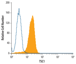
- Detection of TSC1 in Jurkat Human Cell Line by Flow Cytometry. Jurkat human acute T cell leukemia cell line was stained with Human TSC1 Monoclonal Antibody (Catalog # MAB4379, filled histogram) or isotype control antibody (Catalog # MAB0041, open histogram), followed by Phycoerythrin-conjugated Anti-Mouse IgG F(ab')2 Secondary Antibody (Catalog # F0102B). To facilitate intracellular staining, cells were fixed with PFA and permeabilized with saponin.
