Explore
 Validate
 Learn
Learn Western blot
 Immunocytochemistry
Antibody data
- Antibody Data
- Antigen structure
- References [3]
- Comments [0]
- Validations
- Immunocytochemistry [4]
- Other assay [2]
Submit
Validation data
Reference
Comment
Report error
- Product number
- PA5-27366 - Provider product page

- Provider
- Invitrogen Antibodies
- Product name
- EIF2S1 Polyclonal Antibody
- Antibody type
- Polyclonal
- Antigen
- Recombinant full-length protein
- Description
- Recommended positive controls: A549, H1299, HCT-116, rat neuron, HeLa, N2a. Predicted reactivity: Mouse (98%), Rat (98%), Xenopus laevis (92%), Chicken (97%), Rhesus Monkey (100%), Bovine (98%). Store product as a concentrated solution. Centrifuge briefly prior to opening the vial.
- Reactivity
- Human, Mouse, Rat
- Host
- Rabbit
- Isotype
- IgG
- Vial size
- 100 μL
- Concentration
- 0.63 mg/mL
- Storage
- Store at 4°C short term. For long term storage, store at -20°C, avoiding freeze/thaw cycles.
Submitted references Fisetin Protects HaCaT Human Keratinocytes from Fine Particulate Matter (PM(2.5))-Induced Oxidative Stress and Apoptosis by Inhibiting the Endoplasmic Reticulum Stress Response.
Protective Effect of Anthocyanin-Enriched Polyphenols from Hibiscus syriacus L. (Malvaceae) against Ultraviolet B-Induced Damage.
ZNF322A-mediated protein phosphorylation induces autophagosome formation through modulation of IRS1-AKT glucose uptake and HSP-elicited UPR in lung cancer.
Molagoda IMN, Kavinda MHD, Choi YH, Lee H, Kang CH, Lee MH, Lee CM, Kim GY
Antioxidants (Basel, Switzerland) 2021 Sep 18;10(9)
Antioxidants (Basel, Switzerland) 2021 Sep 18;10(9)
Protective Effect of Anthocyanin-Enriched Polyphenols from Hibiscus syriacus L. (Malvaceae) against Ultraviolet B-Induced Damage.
Karunarathne WAHM, Molagoda IMN, Lee KT, Choi YH, Yu SM, Kang CH, Kim GY
Antioxidants (Basel, Switzerland) 2021 Apr 9;10(4)
Antioxidants (Basel, Switzerland) 2021 Apr 9;10(4)
ZNF322A-mediated protein phosphorylation induces autophagosome formation through modulation of IRS1-AKT glucose uptake and HSP-elicited UPR in lung cancer.
Cheung CHY, Hsu CL, Lin TY, Chen WT, Wang YC, Huang HC, Juan HF
Journal of biomedical science 2020 Jun 23;27(1):75
Journal of biomedical science 2020 Jun 23;27(1):75
No comments: Submit comment
Supportive validation
- Submitted by
- Invitrogen Antibodies (provider)
- Main image

- Experimental details
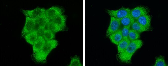
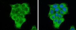
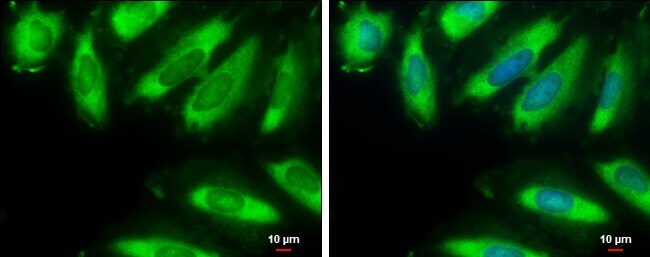
- Immunocytochemistry-Immunofluorescence analysis of eIF2a was performed in HCT 116 cells fixed in 4% paraformaldehyde at RT for 15 min. Green: eIF2a Polyclonal Antibody (Product # PA5-27366) diluted at 1:500. Blue: Hoechst 33342 staining.
- Submitted by
- Invitrogen Antibodies (provider)
- Main image
- Experimental details
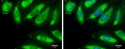
- Immunocytochemistry-Immunofluorescence analysis of eIF2a was performed in HeLa cells fixed in 4% paraformaldehyde at RT for 15 min. Green: eIF2a Polyclonal Antibody (Product # PA5-27366) diluted at 1:5000. Blue: Hoechst 33342 staining. Scale bar = 10 µm.

- Submitted by
- Invitrogen Antibodies (provider)
- Main image
- Experimental details
- Immunocytochemistry-Immunofluorescence analysis of eIF2a was performed in HCT 116 cells fixed in 4% paraformaldehyde at RT for 15 min. Green: eIF2a Polyclonal Antibody (Product # PA5-27366) diluted at 1:500. Blue: Hoechst 33342 staining.

- Submitted by
- Invitrogen Antibodies (provider)
- Main image

- Experimental details
- Immunocytochemistry-Immunofluorescence analysis of eIF2a was performed in HeLa cells fixed in 4% paraformaldehyde at RT for 15 min. Green: eIF2a Polyclonal Antibody (Product # PA5-27366) diluted at 1:5000. Blue: Hoechst 33342 staining. Scale bar = 10 µm.
Supportive validation
- Submitted by
- Invitrogen Antibodies (provider)
- Main image
- Experimental details
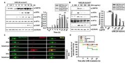
- Figure 8 Effect of anthocyanins from the flower petals of Hibiscus syriacus L. (Malvaceae, AHs) on ER stress and mtROS production. ( A ) and ( B ) The expression of GRP78, ATF4, p-eIF1alpha, CHOP, and beta-actin protein (left) and relative density (right). ( C ) The staining of MitoSOX Red and MitoTracker Green. ( D ) Survival rate of zebrafish. * p < 0.05 and *** p < 0.001 vs. UVB-irradiated cells and ### p < 0.001 vs. untreated cells (UT).

- Submitted by
- Invitrogen Antibodies (provider)
- Main image
- Experimental details
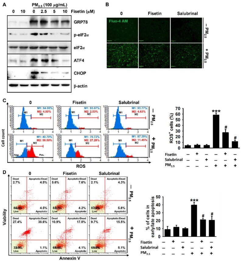
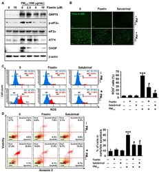
- Figure 4 Fisetin inhibits PM 2.5 -induced apoptosis by alleviating ER stress. HaCaT keratinocytes were treated with fisetin (0-10 uM) for 2 h and subsequently exposed to 100 ug/mL PM 2.5 for 24 h. ( A ) The total proteins were extracted, and Western blotting was performed for detecting the expression of GRP78, p-eIF2alpha, eIF2alpha, ATF4, and CHOP. beta-Actin was used as the loading control. ( B ) The cells were treated with 10 uM fisetin or 20 uM salubrinal in the presence or absence of 100 ug/mL PM 2.5 for 24 h. The cells were incubated with Ca 2+ -sensitive Fluo-4 AM for 10 min and live images were captured using a CELENA S Digital Imaging System. Scale bar = 100 um. ( C , D ) In a parallel experiment, the cells were treated with 10 uM fisetin or 20 uM salubrinal in the presence or absence of 100 ug/mL PM 2.5 for 24 h. The cells were stained with a ( C ) Muse Oxidative Stress Assay Kit and ( D ) Muse Annexin V & Dead Cell Assay Kit. *** p < 0.001 vs. untreated cells and # p < 0.05 vs. PM 2.5 -treated cells.