Explore
 Validate
 Learn
Learn Western blot
Antibody data
- Antibody Data
- Antigen structure
- References [2]
- Comments [0]
- Validations
- Western blot [4]
- Immunohistochemistry [1]
Submit
Validation data
Reference
Comment
Report error
- Product number
- GTX633625 - Provider product page

- Provider
- GeneTex
- Product name
- IDE antibody [GT286]
- Antibody type
- Monoclonal
- Reactivity
- Human, Mouse, Rat
- Host
- Mouse
Submitted references Loss of renal SNX5 results in impaired IDE activity and insulin resistance in mice.
Ablation of retinal ciliopathy protein RPGR results in altered photoreceptor ciliary composition.
Li F, Yang J, Villar VAM, Asico LD, Ma X, Armando I, Sanada H, Yoneda M, Felder RA, Jose PA, Wang X
Diabetologia 2018 Mar;61(3):727-737
Diabetologia 2018 Mar;61(3):727-737
Ablation of retinal ciliopathy protein RPGR results in altered photoreceptor ciliary composition.
Rao KN, Li L, Anand M, Khanna H
Scientific reports 2015 Jun 11;5:11137
Scientific reports 2015 Jun 11;5:11137
No comments: Submit comment
Supportive validation
- Submitted by
- GeneTex (provider)
- Main image
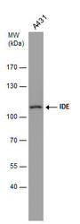
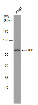
- Experimental details
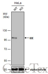
- Whole cell extract (30 ?g) was separated by 7.5% SDS-PAGE, and the membrane was blotted with IDE antibody [GT286] (GTX633625) diluted at 1:1000.
- Submitted by
- GeneTex (provider)
- Main image
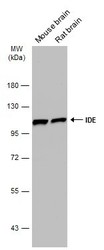
- Experimental details
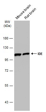
- Various tissue extracts (50 ?g) were separated by 7.5% SDS-PAGE, and the membrane was blotted with IDE antibody [GT286] (GTX633625) diluted at 1:1000. The HRP-conjugated anti-mouse IgG antibody (GTX213111-01) was used to detect the primary antibody.
- Submitted by
- GeneTex (provider)
- Main image
- Experimental details
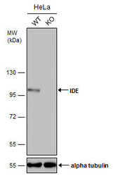
- Wild-type (WT) and IDE knockout (KO) HeLa cell extracts (30 ?g) were separated by 7.5% SDS-PAGE, and the membrane was blotted with IDE antibody [GT286] (GTX633625) diluted at 1:500. The HRP-conjugated anti-mouse IgG antibody (GTX213111-01) was used to detect the primary antibody.
- Submitted by
- GeneTex (provider)
- Main image
- Experimental details
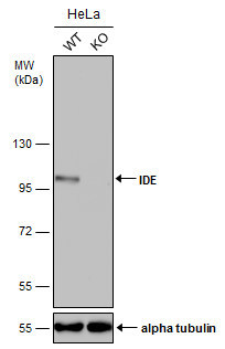
- Wild-type (WT) and IDE knockout (KO) HeLa cell extracts (30 ?g) were separated by 7.5% SDS-PAGE, and the membrane was blotted with IDE antibody [GT286] (GTX633625) diluted at 1:500. The HRP-conjugated anti-mouse IgG antibody (GTX213111-01) was used to detect the primary antibody.

Supportive validation
- Submitted by
- GeneTex (provider)
- Main image
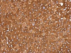
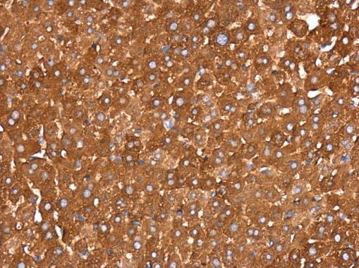
- Experimental details
- IDE antibody [GT286] detects IDE protein at cytoplasm in mouse liver by immunohistochemical analysis. Sample: Paraffin-embedded mouse liver. IDE antibody [GT286] (GTX633625) diluted at 1:200.