Explore
 Validate
 Learn
Learn Western blot
 Immunocytochemistry
Antibody data
- Antibody Data
- Antigen structure
- References [1]
- Comments [0]
- Validations
- Immunocytochemistry [1]
Submit
Validation data
Reference
Comment
Report error
- Product number
- MAB3847 - Provider product page

- Provider
- R&D Systems
- Product name
- Human Notch-4 Intracellular Domain Antibody
- Antibody type
- Monoclonal
- Description
- Protein A or G purified from hybridoma culture supernatant. Detects human Notch-4 Intracellular Domain in direct ELISAs and Western blots. In direct ELISAs and Western blots, no cross-reactivity with recombinant human (rh) Notch-1 Intracellular Domain, rhNotch-2 Intracellular Domain, rhNotch-3, recombinant rat (rr) Notch-1 EC, rrNotch-2 EC, or rrDelta-1 is observed.
- Reactivity
- Human
- Host
- Rat
- Conjugate
- Unconjugated
- Antigen sequence
Q99466- Isotype
- IgG
- Antibody clone number
- 411913
- Vial size
- 100 ug
- Concentration
- LYOPH
- Storage
- Use a manual defrost freezer and avoid repeated freeze-thaw cycles. 12 months from date of receipt, -20 to -70 °C as supplied. 1 month, 2 to 8 °C under sterile conditions after reconstitution. 6 months, -20 to -70 °C under sterile conditions after reconstitution.
Submitted references KSHV-induced notch components render endothelial and mural cell characteristics and cell survival.
Liu R, Li X, Tulpule A, Zhou Y, Scehnet JS, Zhang S, Lee JS, Chaudhary PM, Jung J, Gill PS
Blood 2010 Jan 28;115(4):887-95
Blood 2010 Jan 28;115(4):887-95
No comments: Submit comment
Supportive validation
- Submitted by
- R&D Systems (provider)
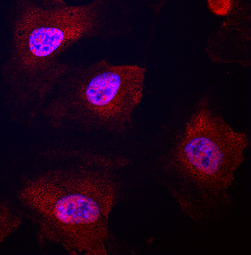
- Main image

- Experimental details
- Notch-4 in Human HUVEC. Notch-4 was detected in immersion fixed HUVEC human umbilical vein endothelial cells using Rat Anti-Human Notch-4 Intracellular Domain Monoclonal Antibody (Catalog # MAB3847) at 10 µg/mL for 3 hours at room temperature. Cells were stained using the NorthernLights™ 557-conjugated Anti-Rat IgG Secondary Antibody (red; Catalog # NL013) and counterstained with DAPI (blue). Specific staining was localized to nuclei and cytoplasm. View our protocol for Fluorescent ICC Staining of Cells on Coverslips.