 Explore
 Validate
 Learn
LearnGTX25484
antibody from GeneTex
Targeting: P4HB
DSI, ERBA2L, GIT, P4Hbeta, PDI, PDIA1, PO4DB, PO4HB, PROHB
 Western blot
 Immunocytochemistry Immunoprecipitation Immunohistochemistry Flow cytometry Blocking/Neutralizing
Antibody data
- Antibody Data
- Antigen structure
- References [0]
- Comments [0]
- Validations
- Western blot [1]
- Immunohistochemistry [1]
- Flow cytometry [3]
Submit
Validation data
Reference
Comment
Report error
- Product number
- GTX25484 - Provider product page

- Provider
- GeneTex
- Proper citation
- GeneTex Cat#GTX25484, RRID:AB_370013
- Product name
- PDI antibody [RL77]
- Antibody type
- Monoclonal
- Reactivity
- Human, Mouse, Rat, Canine, Hamster, Porcine, Simian, Xenopus
- Host
- Mouse
No comments: Submit comment
Supportive validation
- Submitted by
- GeneTex (provider)
- Main image
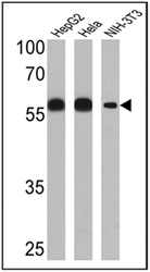
- Experimental details
- Western blot analysis of PDI was performed by loading 25 ug of HepG2 (Lane 1), Hela (Lane 2) and NIH-3T3 (Lane 3) cell lysates onto an SDS polyacrylamide gel. Proteins were transferred to a PVDF membrane and blocked at 4ºC overnight. The membrane was probed with a PDI monoclonal antibody (GTX25484) at a dilution of 1:1000 overnight at 4°C, washed in TBST, and probed with an HRP-conjugated secondary antibody for 1 hr at room temperature in the dark. Chemiluminescent detection was performed. Results show a band at approx. 57 kDa.
- Validation comment
- WB

Supportive validation
- Submitted by
- GeneTex (provider)
- Main image

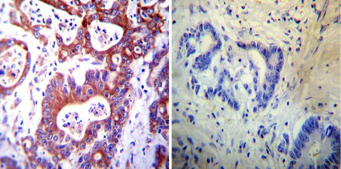
- Experimental details
- Immunohistochemistry was performed on cancer biopsies of deparaffinized human colon carcinoma tissues. To expose target proteins, heat induced antigen retrieval was performed using 10mM sodium citrate (pH6.0) buffer, microwaved for 8-15 minutes. Following antigen retrieval tissues were blocked in 3% BSA-PBS for 30 minutes at room temperature. Tissues were then probed at a dilution of 1:20 with or without PDI antibody [RL77] overnight at 4¢XC in a humidified chamber. Tissues were washed extensively with PBST and endogenous peroxidase activity was quenched with a peroxidase suppressor. Detection was performed using a biotin-conjugated secondary antibody and SA-HRP, followed by colorimetric detection using DAB. Tissues were counterstained with hematoxylin and prepped for mounting.
Supportive validation
- Submitted by
- GeneTex (provider)
- Main image

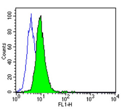
- Experimental details
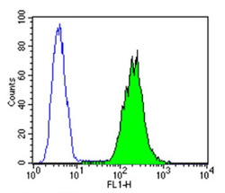
- Flow cytometry analysis of PDI showing weakly positive staining in the cytoplasm of 3T3 cells compared to an isotype control (blue). Cells were harvested, adjusted to a concentration of 1-5x10^6 cells/ml, fixed with 2% paraformaldehyde, washed with PBS, and incubated with PDI monoclonal antibody (GTX25484) at a dilution of 0.5 ug/test for 60 min at room temperature. Cells were then blocked in a solution of 2% BSA-PBS for 30 min at room temperature, incubated for 40 min at room temperature in the dark using a Dylight 488-conjugated goat anti-mouse IgG (H+L) secondary antibody, and re-suspended in PBS for FACS analysis.
- Validation comment
- FACS
- Submitted by
- GeneTex (provider)
- Main image
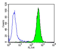
- Experimental details
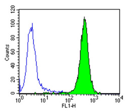
- Flow cytometry analysis of PDI showing positive staining in the cytoplasm of Hela cells compared to an isotype control (blue). Cells were harvested, adjusted to a concentration of 1-5x10^6 cells/ml, fixed with 2% paraformaldehyde, washed with PBS, and incubated with PDI monoclonal antibody (GTX25484) at a dilution of 0.25 ug/test for 60 min at room temperature. Cells were then blocked in a solution of 2% BSA-PBS for 30 min at room temperature, incubated for 40 min at room temperature in the dark using a Dylight 488-conjugated goat anti-mouse IgG (H+L) secondary antibody, and re-suspended in PBS for FACS analysis.
- Validation comment
- FACS
- Submitted by
- GeneTex (provider)
- Main image
- Experimental details
- Flow cytometry analysis of PDI showing positive staining in the cytoplasm of K562 cells compared to an isotype control (blue). Cells were harvested, adjusted to a concentration of 1-5x10^6 cells/ml, fixed with 2% paraformaldehyde, washed with PBS, and incubated with PDI monoclonal antibody at a dilution of 0.25 ug/test for 60 min at room temperature. Cells were then blocked in a solution of 2% BSA-PBS for 30 min at room temperature, incubated for 40 min at room temperature in the dark using a Dylight 488-conjugated goat anti-mouse IgG (H+L) secondary antibody, and re-suspended in PBS for FACS analysis.
- Validation comment
- FACS
