Explore
 Validate
 Learn
LearnA02335-1
antibody from Boster Biological Technology
Targeting: P4HB
DSI, ERBA2L, GIT, P4Hbeta, PDI, PDIA1, PO4DB, PO4HB, PROHB
 Western blot
 ELISA
ELISAAntibody data
- Antibody Data
- Antigen structure
- References [3]
- Comments [0]
- Validations
- Western blot [1]
Submit
Validation data
Reference
Comment
Report error
- Product number
- A02335-1 - Provider product page

- Provider
- Boster Biological Technology
- Product name
- Anti-P4HB Antibody Picoband™
- Antibody type
- Polyclonal
- Description
- Rabbit IgG polyclonal antibody for P4HB detection. Tested with WB, IHC-P, ICC/IF, FCM, Direct ELISA in Human;Monkey;Mouse;Rat.
- Reactivity
- Human, Mouse, Rat, Simian
- Host
- Rabbit
- Vial size
- 100μg/vial
- Concentration
- Add 0.2ml of distilled water will yield a concentration of 500ug/ml.
- Storage
- At -20°C for one year. After reconstitution, at 4°C for one month. It can also be aliquotted and stored frozen at -20°C for a longer time. Avoid repeated freezing and thawing.
- Handling
- Add 0.2ml of distilled water will yield a concentration of 500ug/ml.
Submitted references P4HB, a Novel Hypoxia Target Gene Related to Gastric Cancer Invasion and Metastasis.
ALKBH5 promotes invasion and metastasis of gastric cancer by decreasing methylation of the lncRNA NEAT1.
Prognostic value of hypoxia-inducible factor-1 alpha and prolyl 4-hydroxylase beta polypeptide overexpression in gastric cancer.
Zhang J, Guo S, Wu Y, Zheng ZC, Wang Y, Zhao Y
BioMed research international 2019;2019:9749751
BioMed research international 2019;2019:9749751
ALKBH5 promotes invasion and metastasis of gastric cancer by decreasing methylation of the lncRNA NEAT1.
Zhang J, Guo S, Piao HY, Wang Y, Wu Y, Meng XY, Yang D, Zheng ZC, Zhao Y
Journal of physiology and biochemistry 2019 Aug;75(3):379-389
Journal of physiology and biochemistry 2019 Aug;75(3):379-389
Prognostic value of hypoxia-inducible factor-1 alpha and prolyl 4-hydroxylase beta polypeptide overexpression in gastric cancer.
Zhang J, Wu Y, Lin YH, Guo S, Ning PF, Zheng ZC, Wang Y, Zhao Y
World journal of gastroenterology 2018 Jun 14;24(22):2381-2391
World journal of gastroenterology 2018 Jun 14;24(22):2381-2391
No comments: Submit comment
Supportive validation
- Submitted by
- Boster Biological Technology (provider)
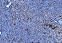
- Main image
- Experimental details
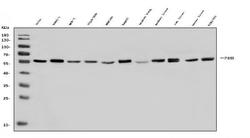
- Western blot analysis of P4HB using anti-P4HB antibody (A02335-1). Electrophoresis was performed on a 5-20% SDS-PAGE gel at 70V (Stacking gel) / 90V (Resolving gel) for 2-3 hours. The sample well of each lane was loaded with 30ug of sample under reducing conditions. Lane 1: human Hela whole cell lysates, Lane 2: human PANC-1 whole cell lysates, Lane 3: human MCF-7 whole cell lysates, Lane 4: human COLO-320 whole cell lysates, Lane 5: human HEK293 whole cell lysates, Lane 6: human HepG2 whole cell lysates, Lane 7: monkey lung tissue lysates, Lane 8: monkey liver tissue lysates, Lane 9: rat liver tissue lysates, Lane 10: mouse liver tissue lysates, Lane 11: mouse NIH/3T3 whole cell lysates. After Electrophoresis, proteins were transferred to a Nitrocellulose membrane at 150mA for 50-90 minutes. Blocked the membrane with 5% Non-fat Milk/ TBS for 1.5 hour at RT. The membrane was incubated with rabbit anti-P4HB antigen affinity purified polyclonal antibody (Catalog # A02335-1) at 0.25 μg/mL overnight at 4°C, then washed with TBS-0.1%Tween 3 times with 5 minutes each and probed with a goat anti-rabbit IgG-HRP secondary antibody at a dilution of 1:5000 for 1.5 hour at RT. The signal is developed using an Enhanced Chemiluminescent detection (ECL) kit (Catalog # EK1002) with Tanon 5200 system. A specific band was detected for P4HB at approximately 57KD. The expected band size for P4HB is at 57KD.
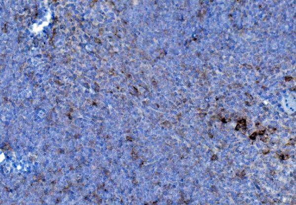
- Additional image