Explore
 Validate
 Learn
Learn Western blot
 Immunocytochemistry
Antibody data
- Antibody Data
- Antigen structure
- References [11]
- Comments [0]
- Validations
- Immunocytochemistry [4]
- Immunohistochemistry [1]
- Flow cytometry [8]
- Other assay [2]
Submit
Validation data
Reference
Comment
Report error
- Product number
- MA3-16537 - Provider product page

- Provider
- Invitrogen Antibodies
- Product name
- MGMT Monoclonal Antibody (MT 23.2)
- Antibody type
- Monoclonal
- Antigen
- Purifed from natural sources
- Description
- Suggested positive control: CEM-CCRF cells. TK6 cells as a negative control. This antibody is mouse reactivity negative.
- Reactivity
- Human, Mouse
- Host
- Mouse
- Isotype
- IgG
- Antibody clone number
- MT 23.2
- Vial size
- 100 μL
- Concentration
- 1 mg/mL
- Storage
- Store at 4°C short term. For long term storage, store at -20°C, avoiding freeze/thaw cycles.
Submitted references Glioma Cells in the Tumor Periphery Have a Stem Cell Phenotype.
Phase 2 study of concurrent radiotherapy and temozolomide followed by temozolomide and lomustine in the treatment of children with high-grade glioma: a report of the Children's Oncology Group ACNS0423 study.
Long-term outcome and MGMT as a predictive marker in 24 patients with atypical pituitary adenomas and pituitary carcinomas given treatment with temozolomide.
MGMT expression and pituitary tumours: relationship to tumour biology.
O6-methylguanine-DNA methyltransferase (MGMT) promoter methylation is a rare event in soft tissue sarcoma.
Hsp27 (HSPB1): a possible surrogate molecular marker for loss of heterozygosity (LOH) of chromosome 1p in oligodendrogliomas but not in astrocytomas.
O6-Methylguanine-DNA methyltransferase protein expression by immunohistochemistry in brain and non-brain systemic tumours: systematic review and meta-analysis of correlation with methylation-specific polymerase chain reaction.
Impact of morphology, MIB-1, p53 and MGMT on outcome in pilocytic astrocytomas.
Salvage therapy with temozolomide in patients with aggressive or metastatic pituitary adenomas: experience in six cases.
Low O6-methylguanine-DNA methyltransferase (MGMT) expression and response to temozolomide in aggressive pituitary tumours.
Prognostic significance of DNA repair proteins MLH1, MSH2 and MGMT expression in non-small-cell lung cancer and precursor lesions.
Munthe S, Petterson SA, Dahlrot RH, Poulsen FR, Hansen S, Kristensen BW
PloS one 2016;11(5):e0155106
PloS one 2016;11(5):e0155106
Phase 2 study of concurrent radiotherapy and temozolomide followed by temozolomide and lomustine in the treatment of children with high-grade glioma: a report of the Children's Oncology Group ACNS0423 study.
Jakacki RI, Cohen KJ, Buxton A, Krailo MD, Burger PC, Rosenblum MK, Brat DJ, Hamilton RL, Eckel SP, Zhou T, Lavey RS, Pollack IF
Neuro-oncology 2016 Oct;18(10):1442-50
Neuro-oncology 2016 Oct;18(10):1442-50
Long-term outcome and MGMT as a predictive marker in 24 patients with atypical pituitary adenomas and pituitary carcinomas given treatment with temozolomide.
Bengtsson D, Schrøder HD, Andersen M, Maiter D, Berinder K, Feldt Rasmussen U, Rasmussen ÅK, Johannsson G, Hoybye C, van der Lely AJ, Petersson M, Ragnarsson O, Burman P
The Journal of clinical endocrinology and metabolism 2015 Apr;100(4):1689-98
The Journal of clinical endocrinology and metabolism 2015 Apr;100(4):1689-98
MGMT expression and pituitary tumours: relationship to tumour biology.
McCormack A, Kaplan W, Gill AJ, Little N, Cook R, Robinson B, Clifton-Bligh R
Pituitary 2013 Jun;16(2):208-19
Pituitary 2013 Jun;16(2):208-19
O6-methylguanine-DNA methyltransferase (MGMT) promoter methylation is a rare event in soft tissue sarcoma.
Jakob J, Hille M, Sauer C, Ströbel P, Wenz F, Hohenberger P
Radiation oncology (London, England) 2012 Oct 30;7:180
Radiation oncology (London, England) 2012 Oct 30;7:180
Hsp27 (HSPB1): a possible surrogate molecular marker for loss of heterozygosity (LOH) of chromosome 1p in oligodendrogliomas but not in astrocytomas.
Castro GN, Cayado-Gutiérrez N, Moncalero VL, Lima P, De Angelis RL, Chávez V, Cuello-Carrión FD, Ciocca DR
Cell stress & chaperones 2012 Nov;17(6):779-90
Cell stress & chaperones 2012 Nov;17(6):779-90
O6-Methylguanine-DNA methyltransferase protein expression by immunohistochemistry in brain and non-brain systemic tumours: systematic review and meta-analysis of correlation with methylation-specific polymerase chain reaction.
Brell M, Ibáñez J, Tortosa A
BMC cancer 2011 Jan 26;11:35
BMC cancer 2011 Jan 26;11:35
Impact of morphology, MIB-1, p53 and MGMT on outcome in pilocytic astrocytomas.
Horbinski C, Hamilton RL, Lovell C, Burnham J, Pollack IF
Brain pathology (Zurich, Switzerland) 2010 May;20(3):581-8
Brain pathology (Zurich, Switzerland) 2010 May;20(3):581-8
Salvage therapy with temozolomide in patients with aggressive or metastatic pituitary adenomas: experience in six cases.
Losa M, Mazza E, Terreni MR, McCormack A, Gill AJ, Motta M, Cangi MG, Talarico A, Mortini P, Reni M
European journal of endocrinology 2010 Dec;163(6):843-51
European journal of endocrinology 2010 Dec;163(6):843-51
Low O6-methylguanine-DNA methyltransferase (MGMT) expression and response to temozolomide in aggressive pituitary tumours.
McCormack AI, McDonald KL, Gill AJ, Clark SJ, Burt MG, Campbell KA, Braund WJ, Little NS, Cook RJ, Grossman AB, Robinson BG, Clifton-Bligh RJ
Clinical endocrinology 2009 Aug;71(2):226-33
Clinical endocrinology 2009 Aug;71(2):226-33
Prognostic significance of DNA repair proteins MLH1, MSH2 and MGMT expression in non-small-cell lung cancer and precursor lesions.
Cooper WA, Kohonen-Corish MR, Chan C, Kwun SY, McCaughan B, Kennedy C, Sutherland RL, Lee CS
Histopathology 2008 Apr;52(5):613-22
Histopathology 2008 Apr;52(5):613-22
No comments: Submit comment
Supportive validation
- Submitted by
- Invitrogen Antibodies (provider)
- Main image
- Experimental details
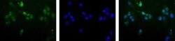
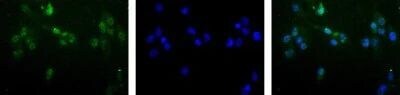
- Immunocytochemistry analysis of MGMT in HeLa cells. Samples were incubated in MGMT monoclonal antibody (Product # MA3-16537). MGMT (Green). Nuclei (Blue) are counterstained using Hoechst 33258..

- Submitted by
- Invitrogen Antibodies (provider)
- Main image
- Experimental details
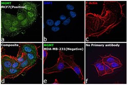
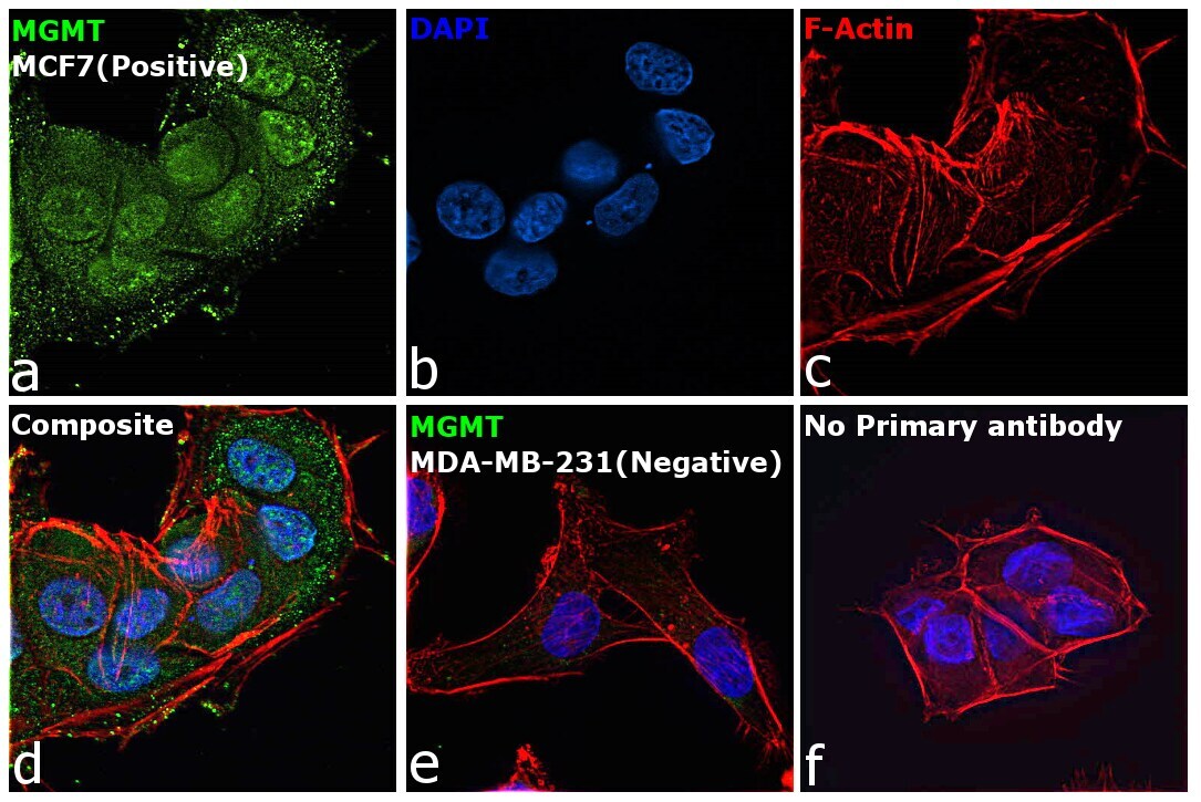
- Immunofluorescence analysis of Methylated-DNA-protein-cysteine methyltransferase was performed using 70% confluent log phase MCF7 cells. The cells were fixed with 4% paraformaldehyde for 10 minutes, permeabilized with 0.1% Triton™ X-100 for 15 minutes, and blocked with 2% BSA for 45 minutes at room temperature. The cells were labeled with MGMT Monoclonal Antibody (MT 23.2) (Product # MA3-16537) at 1:500 dilution in 0.1% BSA, incubated at 4 degree celsius overnight and then labeled with Goat anti-Mouse IgG (H+L) Highly Cross-Adsorbed Secondary Antibody, Alexa Fluor Plus 488 (Product # A32723), (1:2000 dilution), for 45 minutes at room temperature (Panel a: Green). Nuclei (Panel b:Blue) were stained with ProLong™ Diamond Antifade Mountant with DAPI (Product # P36962). F-actin (Panel c: Red) was stained with Rhodamine Phalloidin (Product # R415, 1:300 dilution). Panel d represents the merged image showing nucleus and cytoplasm localization. Panel e represents MDA-MB-231 cells which is negative for MGMT. Panel f represents control cells with no primary antibody to assess background. The images were captured at 60X magnification.

- Submitted by
- Invitrogen Antibodies (provider)
- Main image

- Experimental details
- Immunofluorescence analysis of Methylated-DNA-protein-cysteine methyltransferase was performed using 70% confluent log phase MCF7 cells. The cells were fixed with 4% paraformaldehyde for 10 minutes, permeabilized with 0.1% Triton™ X-100 for 15 minutes, and blocked with 2% BSA for 45 minutes at room temperature. The cells were labeled with MGMT Monoclonal Antibody (MT 23.2) (Product # MA3-16537) at 1:500 dilution in 0.1% BSA, incubated at 4 degree celsius overnight and then labeled with Goat anti-Mouse IgG (H+L) Highly Cross-Adsorbed Secondary Antibody, Alexa Fluor Plus 488 (Product # A32723), (1:2000 dilution), for 45 minutes at room temperature (Panel a: Green). Nuclei (Panel b:Blue) were stained with ProLong™ Diamond Antifade Mountant with DAPI (Product # P36962). F-actin (Panel c: Red) was stained with Rhodamine Phalloidin (Product # R415, 1:300 dilution). Panel d represents the merged image showing nucleus and cytoplasm localization. Panel e represents MDA-MB-231 cells which is negative for MGMT. Panel f represents control cells with no primary antibody to assess background. The images were captured at 60X magnification.
- Submitted by
- Invitrogen Antibodies (provider)
- Main image

- Experimental details
- Immunocytochemistry analysis of MGMT in HeLa cells. Samples were incubated in MGMT monoclonal antibody (Product # MA3-16537). MGMT (Green). Nuclei (Blue) are counterstained using Hoechst 33258..
Supportive validation
- Submitted by
- Invitrogen Antibodies (provider)
- Main image
- Experimental details
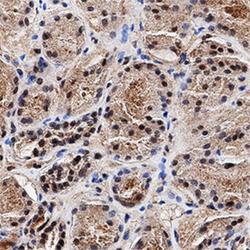
- Immunohistochemical analysis of MGMT in formalin fixed paraffin-embedded (FFPE) human kidney. Samples were incubated in MGMT monoclonal antibody (Product # MA3-16537) using a dilution of 1:2000. Bond Rx autostainer (Leica Biosystems). The assay involved 20 minutes of heat induced antigen retrieval (HIER) using 10mM sodium citrate buffer (pH 6.0) and endogenous peroxidase quenching with peroxide block. The sections were incubated with primary antibody for 30 minutes and Bond Polymer Refine Detection (Leica Biosystems) with DAB was used for signal development followed by counterstaining with hematoxylin. Whole slide scanning and capturing of representative images was performed using Aperio AT2 (Leica Biosystems). Nuclear staining of MGMT was observed. Staining was performed by Histowiz.

Supportive validation
- Submitted by
- Invitrogen Antibodies (provider)
- Main image
- Experimental details
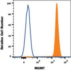
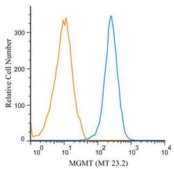
- Flow cytometry of MGMT in Human Jurkat cell line. Samples were incubated in MGMT monoclonal antibody (Product # MA3-16537) or Mouse IgG2B isotype control followed by APC-conjugated Anti-Mouse IgG Secondary Antibody. To facilitate intracellular staining, cells were fixed with FlowX FoxP3 Fixation & Permeabilization Buffer Kit .

- Submitted by
- Invitrogen Antibodies (provider)
- Main image
- Experimental details
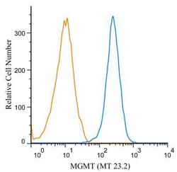
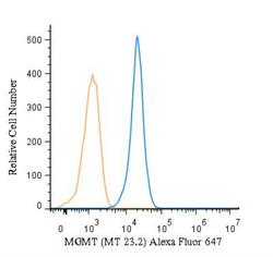
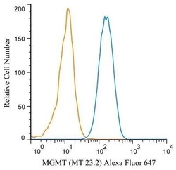
- Flow cytometry of MGMT in Jurkat cells. Samples were incubated in MGMT monoclonal antibody (Product # MA3-16537) using a dilution of 1 µg/mL for 30 minutes at room temperature followed by mouse F(ab)2 IgG (H+L) APC-conjugated secondary antibody. Antibody (blue) and a matched isotype control (orange). Cells were fixed with 4% PFA and then permeablized with 0.1% saponin.
- Submitted by
- Invitrogen Antibodies (provider)
- Main image

- Experimental details
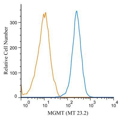
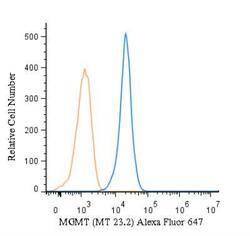
- Flow cytometry of MGMT in Raji cells. Samples were incubated in MGMT monoclonal antibody (Product # MA3-16537) using a dilution of 2 µg/mL for 30 minutes at room temperature. Antibody (blue) and a matched isotype control (orange). Cells were fixed with 4% PFA and then permeablized with 0.1% saponin. Both antibodies were conjugated to Alexa Fluor 647.

- Submitted by
- Invitrogen Antibodies (provider)
- Main image
- Experimental details
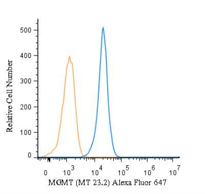
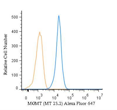
- Flow cytometry of MGMT in Jurkat cells. Samples were incubated in MGMT monoclonal (Product # MA3-16537) using a dilution of 2.5 µg/mL for 30 minutes at room temperature. Antibody (blue) and a matched isotype control (orange). Cells were fixed with 4% PFA and then permeablized with 0.1% saponin. Both antibodies were conjugated to Alexa Fluor 647.
- Submitted by
- Invitrogen Antibodies (provider)
- Main image

- Experimental details
- Flow cytometry of MGMT in Human Jurkat cell line. Samples were incubated in MGMT monoclonal antibody (Product # MA3-16537) or Mouse IgG2B isotype control followed by APC-conjugated Anti-Mouse IgG Secondary Antibody. To facilitate intracellular staining, cells were fixed with FlowX FoxP3 Fixation & Permeabilization Buffer Kit .

- Submitted by
- Invitrogen Antibodies (provider)
- Main image
- Experimental details
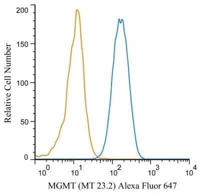
- Flow cytometry of MGMT in Jurkat cells. Samples were incubated in MGMT monoclonal antibody (Product # MA3-16537) using a dilution of 1 µg/mL for 30 minutes at room temperature followed by mouse F(ab)2 IgG (H+L) APC-conjugated secondary antibody. Antibody (blue) and a matched isotype control (orange). Cells were fixed with 4% PFA and then permeablized with 0.1% saponin.

- Submitted by
- Invitrogen Antibodies (provider)
- Main image
- Experimental details
- Flow cytometry of MGMT in Raji cells. Samples were incubated in MGMT monoclonal antibody (Product # MA3-16537) using a dilution of 2 µg/mL for 30 minutes at room temperature. Antibody (blue) and a matched isotype control (orange). Cells were fixed with 4% PFA and then permeablized with 0.1% saponin. Both antibodies were conjugated to Alexa Fluor 647.
- Submitted by
- Invitrogen Antibodies (provider)
- Main image
- Experimental details
- Flow cytometry of MGMT in Jurkat cells. Samples were incubated in MGMT monoclonal (Product # MA3-16537) using a dilution of 2.5 µg/mL for 30 minutes at room temperature. Antibody (blue) and a matched isotype control (orange). Cells were fixed with 4% PFA and then permeablized with 0.1% saponin. Both antibodies were conjugated to Alexa Fluor 647.
Supportive validation
- Submitted by
- Invitrogen Antibodies (provider)
- Main image

- Experimental details
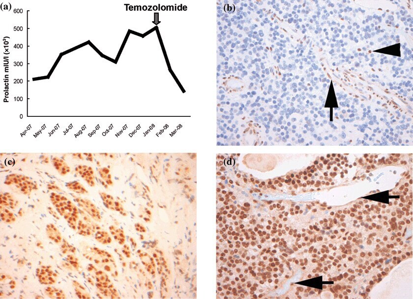
- 1 (a) Serum prolactin concentrations in case 1 before and after commencement of temozolomide therapy (arrowed). (b) MGMT immunohistochemistry (IHC): case 1 pituitary tumour shows completely negative nuclear staining for MGMT. The endothelial cells (arrow) and lymphocytes (arrowhead) act as internal positive controls (original magnification 400x). (c) MGMT IHC: case 1 adjacent non-neoplastic pituitary tissue shows strong nuclear staining for MGMT. Cytoplasmic staining is considered nonspecific (original magnification 400x). (d) MGMT IHC: case 2 pituitary tumour shows diffuse strong nuclear staining for MGMT with positive staining in endothelial cells (arrows) (original magnification 400x).
- Submitted by
- Invitrogen Antibodies (provider)
- Main image

- Experimental details
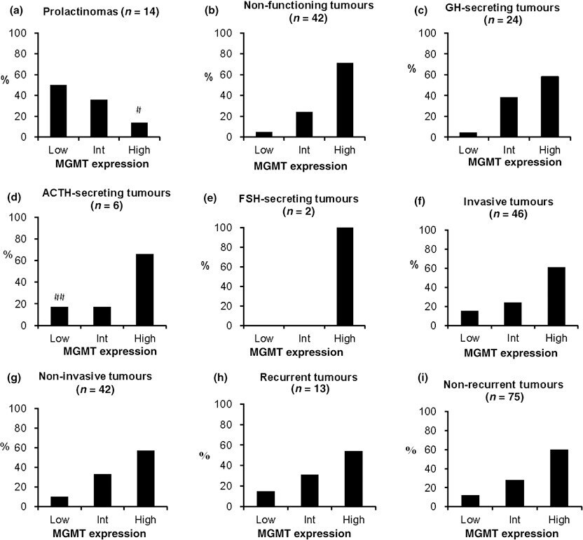
- 2 MGMT immunohistochemistry: (a) prolactinomas; (b) non-functioning pituitary tumours; (c) GH-secreting tumours; (d) ACTH-secreting tumours; (e) FSH-secreting tumours; (f) invasive tumours; (g) noninvasive tumours; (h) recurrent tumours; (i) nonrecurrent tumours. Prolactinomas vs. other pituitary tumour subtypes, P < 0*001; invasive vs. noninvasive tumours, P = 0*553; recurrent vs. nonrecurrent tumours, P = 0*75. # Includes a malignant prolactinoma. ## Includes a malignant ACTH-secreting tumour.