Explore
 Validate
 Learn
Learn Western blot
Antibody data
- Antibody Data
- Antigen structure
- References [3]
- Comments [0]
- Validations
- Western blot [13]
- Immunocytochemistry [3]
- Immunohistochemistry [1]
Submit
Validation data
Reference
Comment
Report error
- Product number
- GTX109832 - Provider product page

- Provider
- GeneTex
- Proper citation
- GeneTex Cat#GTX109832, RRID:AB_1952426
- Product name
- alpha Tubulin 1A antibody
- Antibody type
- Polyclonal
- Reactivity
- Human, Mouse, Rat, Drosophila
- Host
- Rabbit
Submitted references Role of estrogen receptors and Src signaling in mechanisms of bone metastasis by estrogen receptor positive breast cancers.
Aryl hydrocarbon receptor in association with RelA modulates IL-6 expression in non-smoking lung cancer.
Translocation of Helicobacter pylori CagA into Human B lymphocytes, the origin of mucosa-associated lymphoid tissue lymphoma.
Chiu JH, Wen CS, Wang JY, Hsu CY, Tsai YF, Hung SC, Tseng LM, Shyr YM
Journal of translational medicine 2017 May 4;15(1):97
Journal of translational medicine 2017 May 4;15(1):97
Aryl hydrocarbon receptor in association with RelA modulates IL-6 expression in non-smoking lung cancer.
Chen PH, Chang H, Chang JT, Lin P
Oncogene 2012 May 17;31(20):2555-65
Oncogene 2012 May 17;31(20):2555-65
Translocation of Helicobacter pylori CagA into Human B lymphocytes, the origin of mucosa-associated lymphoid tissue lymphoma.
Lin WC, Tsai HF, Kuo SH, Wu MS, Lin CW, Hsu PI, Cheng AL, Hsu PN
Cancer research 2010 Jul 15;70(14):5740-8
Cancer research 2010 Jul 15;70(14):5740-8
No comments: Submit comment
Supportive validation
- Submitted by
- GeneTex (provider)
- Main image
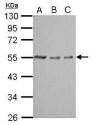
- Experimental details
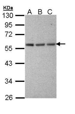
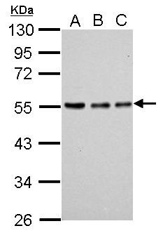
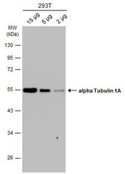
- Sample (whole cell lysate) A: 293T 20ug B: 293T 10ug C: 293T 5ug 10% SDS PAGE GTX109832 diluted at 1:10000
- Validation comment
- WB

- Submitted by
- GeneTex (provider)
- Main image

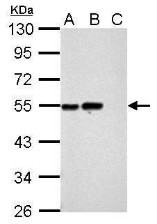
- Experimental details
- Sample (30 ug of cell lysate) A: HeLa B: HeLa cytosol fraction C: HeLa nucleus fraction 7.5% SDS PAGE GTX109832 diluted at 1:10000
- Validation comment
- WB
- Submitted by
- GeneTex (provider)
- Main image
- Experimental details
- Sample (30 ug of whole cell lysate) A: NIH-3T3 10% SDS PAGE GTX109832 diluted at 1:10000
- Validation comment
- WB
- Submitted by
- GeneTex (provider)
- Main image

- Experimental details
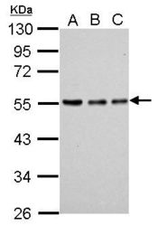
- Sample (30 ug of whole cell lysate)A: HeLaB: Hep G2 (GTX27900)C: Molt-4 (GTX27912)10% SDS PAGEGTX109832 diluted at 1:10000
- Validation comment
- WB
- Submitted by
- GeneTex (provider)
- Main image
- Experimental details
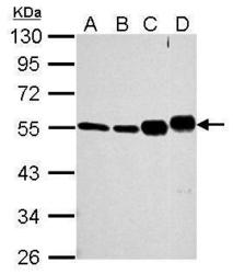
- Sample (30 ug of whole cell lysate) A: 293T B: NIH-3T3 C: Mouse brain D: Rat brain 7.5% SDS PAGE GTX109832 diluted at 1:10000
- Validation comment
- WB

- Submitted by
- GeneTex (provider)
- Main image
- Experimental details
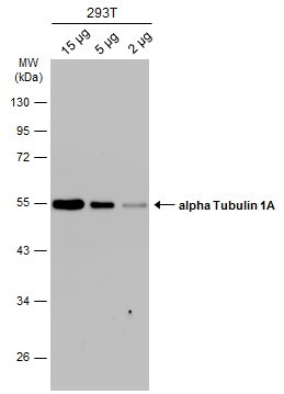
- Sample (whole cell lysate) A: 293T 20ug B: 293T 10ug C: 293T 5ug 10% SDS PAGE GTX109832 diluted at 1:10000 The HRP-conjugated anti-rabbit IgG antibody (GTX213110-01) was used to detect the primary antibody.
- Submitted by
- GeneTex (provider)
- Main image
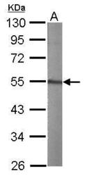
- Experimental details
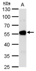
- alpha Tubulin 1A antibody detects alpha Tubulin 1A protein by western blot analysis.A. 50 ?g rat brain lysate/extract10 % SDS-PAGEalpha Tubulin 1A antibody (GTX109832) dilution: 1:10000
- Validation comment
- WB

- Submitted by
- GeneTex (provider)
- Main image
- Experimental details
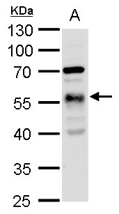
- alpha Tubulin 1A antibody detects alpha Tubulin 1A protein by western blot analysis.A. 30 ?g drosophila lysate/extract10 % SDS-PAGEalpha Tubulin 1A antibody (GTX109832) dilution: 1:1000
- Validation comment
- WB

- Submitted by
- GeneTex (provider)
- Main image

- Experimental details
- alpha Tubulin 1A antibody detects alpha Tubulin 1A protein by western blot analysis.A. 30 ?g drosophila lysate/extract10% SDS-PAGEalpha Tubulin 1A antibody (GTX109832) dilution: 1:1000The HRP-conjugated anti-rabbit IgG antibody (GTX213110-01) was used to detect the primary antibody.
- Submitted by
- GeneTex (provider)
- Main image
- Experimental details
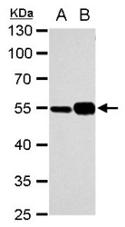
- alpha Tubulin 1A antibody detects alpha Tubulin 1A protein by western blot analysis.A. 30 ?g NIH-3T3 whole cell extract B. 30 ?g mouse brain extract10% SDS-PAGEalpha Tubulin 1A antibody (GTX109832) dilution: 1:10000 The HRP-conjugated anti-rabbit IgG antibody (GTX213110-01) was used to detect the primary antibody.

- Submitted by
- GeneTex (provider)
- Main image

- Experimental details
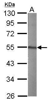
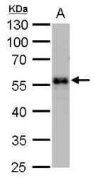
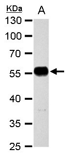
- alpha Tubulin 1A antibody detects alpha Tubulin 1A protein by western blot analysis.A. 30 ?g rat brain extract10% SDS-PAGEalpha Tubulin 1A antibody (GTX109832) dilution: 1:10000 The HRP-conjugated anti-rabbit IgG antibody (GTX213110-01) was used to detect the primary antibody.
- Submitted by
- GeneTex (provider)
- Main image

- Experimental details
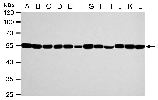
- alpha Tubulin 1A antibody detects alpha Tubulin 1A protein by western blot analysis.A. 30 ?g Jurkat whole cell extractB. 30 ?g Raji whole cell extractC. 30 ?g 293T whole cell extractD. 30 ?g A431 whole cell extractE. 30 ?g HeLa whole cell extractF. 30 ?g HepG2 whole cell extractG. 30 ?g H1299 whole cell extractH. 30 ?g HCT116 whole cell extractI. 30 ?g MCF-7 whole cell extractJ. 30 ?g NT2D1 whole cell extractK. 30 ?g PC-3 whole cell extractL. 30 ?g U87-MG whole cell extract10% SDS-PAGEalpha Tubulin 1A antibody (GTX109832) dilution: 1:10000 The HRP-conjugated anti-rabbit IgG antibody (GTX213110-01) was used to detect the primary antibody.
- Submitted by
- GeneTex (provider)
- Main image
- Experimental details
- Various whole cell extracts were separated by 10% SDS-PAGE, and the membrane was blotted with alpha Tubulin 1A antibody (GTX109832) diluted at 1:10000. The HRP-conjugated anti-rabbit IgG antibody (GTX213110-01) was used to detect the primary antibody.
Supportive validation
- Submitted by
- GeneTex (provider)
- Main image
- Experimental details
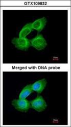
- Immunofluorescence analysis of paraformaldehyde-fixed A431, using Tubulin alpha 1A(GTX109832) antibody at 1:200 dilution.

- Submitted by
- GeneTex (provider)
- Main image
- Experimental details
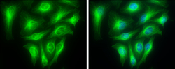
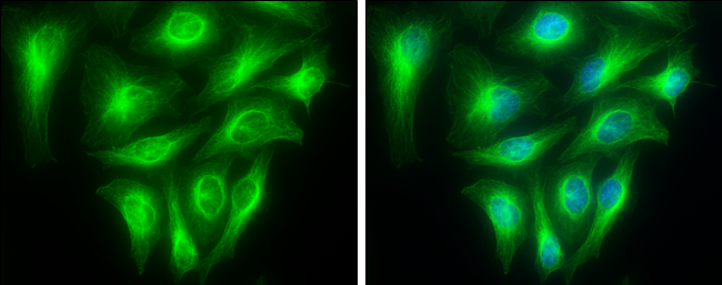
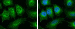
- alpha Tubulin 1A antibody detects alpha Tubulin 1A protein at cytoskeleton by immunofluorescent analysis.Sample: HeLa cells were fixed in 4% paraformaldehyde at RT for 15 min.Green: alpha Tubulin 1A stained by alpha Tubulin 1A antibody (GTX109832) diluted at 1:500.Blue: Hoechst 33342 staining.
- Submitted by
- GeneTex (provider)
- Main image
- Experimental details
- alpha Tubulin 1A antibody detects alpha Tubulin 1A protein at cytoskeleton by immunofluorescent analysis.Sample: HeLa cells were fixed in 4% paraformaldehyde at RT for 15 min.Green: alpha Tubulin 1A stained by alpha Tubulin 1A antibody (GTX109832) diluted at 1:500.Blue: Hoechst 33342 staining.

Supportive validation
- Submitted by
- GeneTex (provider)
- Main image
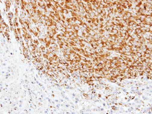
- Experimental details
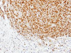
- Immunohistochemical analysis of paraffin-embedded SAS xenograft, using alpha Tubulin 1A(GTX109832) antibody at 1:500 dilution.