Explore
 Validate
 Learn
Learn Western blot
 Immunocytochemistry
Antibody data
- Antibody Data
- Antigen structure
- References [7]
- Comments [0]
- Validations
- Immunocytochemistry [7]
- Immunohistochemistry [1]
- Other assay [4]
Submit
Validation data
Reference
Comment
Report error
- Product number
- PA5-22060 - Provider product page

- Provider
- Invitrogen Antibodies
- Product name
- TUBA1A Polyclonal Antibody
- Antibody type
- Polyclonal
- Antigen
- Recombinant full-length protein
- Description
- Recommended positive controls: Jurkat, Raji, 293T, A431, HeLa, HepG2, H1299, HCT116, MCF-7, NT2D1, PC-3, U87-MG, NIH-3T3, mouse brain, rat brain, Drosophila. Predicted reactivity: Mouse (100%), Rat (100%), Zebrafish (100%), Xenopus laevis (98%), Pig (99%), Rhesus Monkey (100%), Chimpanzee (100%), Bovine (100%). Store product as a concentrated solution. Centrifuge briefly prior to opening the vial.
- Reactivity
- Human, Mouse, Rat, Drosophila
- Host
- Rabbit
- Isotype
- IgG
- Vial size
- 100 μL
- Concentration
- 1.14 mg/mL
- Storage
- Store at 4°C short term. For long term storage, store at -20°C, avoiding freeze/thaw cycles.
Submitted references Absence of detectable SARS-CoV-2 replication in ex vivo cultured cornea and cornea-derived epithelial cells.
Antibody RING-Mediated Destruction of Endogenous Proteins.
Does butein affect adipogenesis?
Hepatoprotective Activity of InlB321/15, the HGFR Ligand of Bacterial Origin, in CCI4-Induced Acute Liver Injury Mice.
Differential miRNA expression profiling reveals miR-205-3p to be a potential radiosensitizer for low- dose ionizing radiation in DLD-1 cells.
Iron importers Zip8 and Zip14 are expressed in retina and regulated by retinal iron levels.
The Gustatory Signaling Pathway and Bitter Taste Receptors Affect the Development of Obesity and Adipocyte Metabolism in Mice.
Bayyoud T, Vavouras Syrigos G, Ruetalo Buschinger N, Wude J, Businger R, Hu D, Iftner A, Thaler S, Schindler M
Graefe's archive for clinical and experimental ophthalmology = Albrecht von Graefes Archiv fur klinische und experimentelle Ophthalmologie 2023 Feb;261(2):435-446
Graefe's archive for clinical and experimental ophthalmology = Albrecht von Graefes Archiv fur klinische und experimentelle Ophthalmologie 2023 Feb;261(2):435-446
Antibody RING-Mediated Destruction of Endogenous Proteins.
Ibrahim AFM, Shen L, Tatham MH, Dickerson D, Prescott AR, Abidi N, Xirodimas DP, Hay RT
Molecular cell 2020 Jul 2;79(1):155-166.e9
Molecular cell 2020 Jul 2;79(1):155-166.e9
Does butein affect adipogenesis?
Hemmeryckx B, Vranckx C, Bauters D, Lijnen HR, Scroyen I
Adipocyte 2019 Dec;8(1):209-222
Adipocyte 2019 Dec;8(1):209-222
Hepatoprotective Activity of InlB321/15, the HGFR Ligand of Bacterial Origin, in CCI4-Induced Acute Liver Injury Mice.
Chalenko Y, Sobyanin K, Sysolyatina E, Midiber K, Kalinin E, Lavrikova A, Mikhaleva L, Ermolaeva S
Biomedicines 2019 Apr 11;7(2)
Biomedicines 2019 Apr 11;7(2)
Differential miRNA expression profiling reveals miR-205-3p to be a potential radiosensitizer for low- dose ionizing radiation in DLD-1 cells.
Andaur R, Tapia JC, Moreno J, Soto L, Armisen R, Marcelain K
Oncotarget 2018 May 29;9(41):26387-26405
Oncotarget 2018 May 29;9(41):26387-26405
Iron importers Zip8 and Zip14 are expressed in retina and regulated by retinal iron levels.
Sterling J, Guttha S, Song Y, Song D, Hadziahmetovic M, Dunaief JL
Experimental eye research 2017 Feb;155:15-23
Experimental eye research 2017 Feb;155:15-23
The Gustatory Signaling Pathway and Bitter Taste Receptors Affect the Development of Obesity and Adipocyte Metabolism in Mice.
Avau B, Bauters D, Steensels S, Vancleef L, Laermans J, Lesuisse J, Buyse J, Lijnen HR, Tack J, Depoortere I
PloS one 2015;10(12):e0145538
PloS one 2015;10(12):e0145538
No comments: Submit comment
Supportive validation
- Submitted by
- Invitrogen Antibodies (provider)
- Main image
- Experimental details
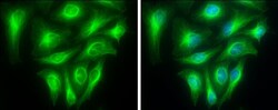
- Immunofluorescent analysis of Tubulin alpha 1A in paraformaldehyde-fixed A431 cells using a Tubulin alpha 1A polyclonal antibody (Product # PA5-22060) at a 1:200 dilution.

- Submitted by
- Invitrogen Antibodies (provider)
- Main image

- Experimental details
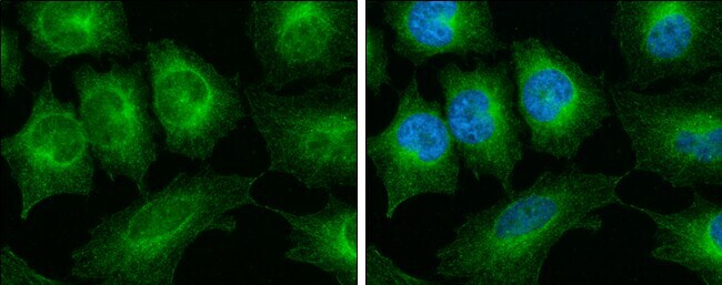
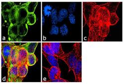
- Immunofluorescence analysis of alpha Tubulin antibody was performed using 70% confluent log phase LNCaP cells. The cells were fixed with 4% paraformaldehyde for 10 minutes, permeabilized with 0.1% Triton™ X-100 for 10 minutes, and blocked with 1% BSA for 1 hour at room temperature. The cells were labeled with Alpha Tubulin Rabbit Polyclonal Antibody (Product # PA5-22060) at 2 µg/mL in 0.1% BSA and incubated for 3 hours at room temperature and then labeled with Goat anti-Rabbit IgG (H+L) Superclonal™ Secondary Antibody, Alexa Fluor® 488 conjugate (Product # A27034) at a dilution of 1:2000 for 45 minutes at room temperature (Panel a: green). Nuclei (Panel b: blue) were stained with SlowFade® Gold Antifade Mountant with DAPI (Product # S36938). F-actin (Panel c: red) was stained with Rhodamine Phalloidin (Product # R415, 1:300). Panel d represents the merged image showing cytoplasmic localization. Panel e shows the no primary antibody control. The images were captured at 60X magnification.

- Submitted by
- Invitrogen Antibodies (provider)
- Main image

- Experimental details
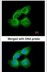
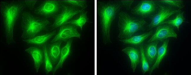
- Immunocytochemistry-Immunofluorescence analysis of alpha Tubulin was performed in HeLa cells fixed in 4% paraformaldehyde at RT for 15 min. Green: alpha Tubulin Polyclonal Antibody (Product # PA5 22060) diluted at 1:500. Blue: Hoechst 33342 staining.

- Submitted by
- Invitrogen Antibodies (provider)
- Main image

- Experimental details
- Immunocytochemistry-Immunofluorescence analysis of alpha Tubulin was performed in HeLa cells fixed in 4% paraformaldehyde at RT for 15 min. Green: alpha Tubulin Polyclonal Antibody (Product # PA5 22060) diluted at 1:500. Blue: Hoechst 33342 staining.
- Submitted by
- Invitrogen Antibodies (provider)
- Main image
- Experimental details
- Immunofluorescence analysis of alpha Tubulin antibody was performed using 70% confluent log phase LNCaP cells. The cells were fixed with 4% paraformaldehyde for 10 minutes, permeabilized with 0.1% Triton™ X-100 for 10 minutes, and blocked with 1% BSA for 1 hour at room temperature. The cells were labeled with Alpha Tubulin Rabbit Polyclonal Antibody (Product # PA5-22060) at 2 µg/mL in 0.1% BSA and incubated for 3 hours at room temperature and then labeled with Goat anti-Rabbit IgG (Heavy Chain) Superclonal™ Secondary Antibody, Alexa Fluor® 488 conjugate (Product # A27034) at a dilution of 1:2000 for 45 minutes at room temperature (Panel a: green). Nuclei (Panel b: blue) were stained with SlowFade® Gold Antifade Mountant with DAPI (Product # S36938). F-actin (Panel c: red) was stained with Rhodamine Phalloidin (Product # R415, 1:300). Panel d represents the merged image showing cytoplasmic localization. Panel e shows the no primary antibody control. The images were captured at 60X magnification.

Supportive validation
- Submitted by
- Invitrogen Antibodies (provider)
- Main image
- Experimental details
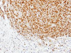
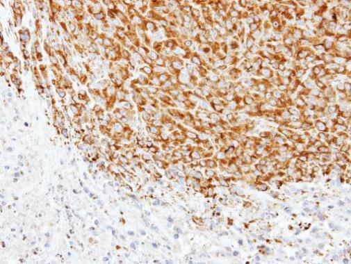
- Immunohistochemical analysis of paraffin-embedded SAS xenograft, using alpha Tubulin 1A (Product # PA5-22060) antibody at 1:500 dilution. Antigen Retrieval: EDTA based buffer, pH 8.0, 15 min.
Supportive validation
- Submitted by
- Invitrogen Antibodies (provider)
- Main image

- Experimental details
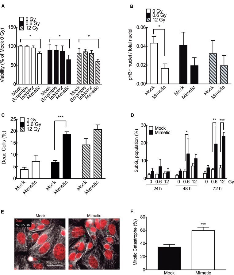
- Figure 7 miR-205-3p increases radiosensitivity at low doses of radiation ( A ) DLD-1 cells were mock-transfected or transfected with a scrambled miR control, an anti-miR-205-3p (inhibitor) or a miR-205-3p mimic. Viability was evaluated 48 h post irradiation with 0, 0.6 or 12 Gy, by MTS assay. Results are expressed as a percentage of mock-transfected non-irradiated cells (0 Gy). ( B ) Proliferation (mitotic index) was determined by quantifying cells with positive staining for phosphor-histone H3 in mock and mimetic transfected cells, 48 hours after irradiation. ( C ) Cell death was evaluated by trypan blue exclusion assay in mock and mimetic transfected cells, 48 hours after irradiation. Data represent percentage of cell death in each condition. ( D ) Effect of miR-205-3p on apoptosis was evaluated by quantification of subG1 population in mock and mimetic transfected cells, 24, 48 and 72 hours after irradiation. ( E ) Evaluation of nuclear morphology of mock and mimetic transfected DLD-1 cells, 72 hours after irradiation (12 Gy). Nuclei were stained with DAPI (Red) and cytoplasm with alpha-tubulin (white). Green head arrows points to fragmented nuclei, features typical of mitotic catastrophe. Scale bar: 50 mum. ( F ) Cells with mitotic catastrophe morphology were counted in 5 different fields and normalized to the number of cells. At least 100 cells were evaluated per sample. Data represent the means +- S.D. of at least 3 independent experiments. * P < 0.05; ** P < 0.0
- Submitted by
- Invitrogen Antibodies (provider)
- Main image
- Experimental details
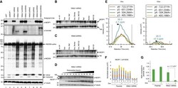
- Figure 4 Degradation of Endogenous NEDD8 Protease NEDP1 with ARMeD Constructs (A) HeLa Flp-in/T.Rex cells were transfected with non-targeting (siNT, lane 1) or NEDP1 (siNEDP1, lane 2) siRNA, and cell extracts harvested 72 h after transfection. Lanes 3-10: HeLa Flp-in/T.Rex cells engineered to inducibly express NEDP1 specific nanobody-RING constructs were untreated (-) or doxycycline-treated (+) for 24 h. Protein levels were analyzed by western blotting using anti-NEDP1, anti-camelid, and anti-NEDD8 antibodies. alpha-Tubulin was used as loading control. NEDD8-cullins and NEDD8 monomers and dimers are indicated by arrows. (B and C) To establish the pathway of protein degradation by NNb2-1xRING (B) and NNb9-2xRING (C), cells were incubated with autophagy inhibitor bafilomycin A1 (Baf, 100 nM) or proteasome inhibitors bortezomib (1 muM) or MG132 (10 mug/mL) for 1.5 h prior to 16 h doxycycline induction. The role of other E3 ligases in degradation of substrate was examined by subjecting cells to inhibitors without Dox induction. (D) Induction of NEDP1 ARMeD fusions and rate of NEDP1 degradation after doxycycline addition was assessed by western blotting using anti-NEDP1 and anti-camelid antibodies. A non-specific (NS) band recognized by the NEDP1 antibody served as an additional loading control. (E) Parallel reaction monitoring to quantify NEDP1 depletion. Example MS2 chromatograms for fragment ions y3-y6 of the NEDP1 peptide LAFVEEK with and without doxycycline treatment. Dashed

- Submitted by
- Invitrogen Antibodies (provider)
- Main image
- Experimental details
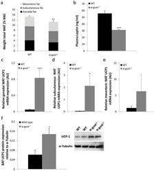
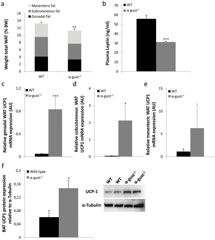
- Fig 2 Difference in adiposity between high-fat diet (19 weeks) induced obese WT and alpha-gust -/- mice. (a) The sum of gonadal, subcutaneous and mesenteric white adipose tissue mass as percentage of total body weight in WT (n = 16) and alpha-gust -/- mice (n = 16). (b) Plasma leptin levels in WT (n = 10) and alpha-gust -/- mice (n = 11). (c-e) Relative gonadal, subcutaneous and mesenteric fat UCP1 mRNA levels in WT (n = 6-9) and alpha-gust -/- mice (n = 6-9). (f) Relative intrascapular brown adipose tissue UCP1 protein level expression in WT (n = 4) and alpha-gust -/- mice (n = 4) as determined by Western blot. *: P
- Submitted by
- Invitrogen Antibodies (provider)
- Main image
- Experimental details
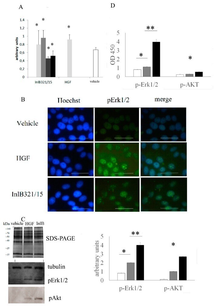
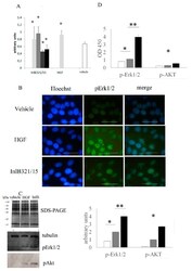
- Figure 1 InlB321/15 activates Erk1/2 and Akt-controlled signaling pathways and provides a mitogenic effect in vitro. ( A ) MTT testing of HepG2 cell survival and proliferation. Cells were grown for 72 h with increasing InlB321/15 concentrations (100, 250, 500, or 1000 ng*mL -1 ) or rhHGF (100 ng*mL -1 , as recommended by a manufacturer); ( B ) distribution of phospho-Erk1/2 in cells. Cells were treated with InlB321/15 or rhHGF for 15 min, then labelled with Hoechst to label nuclei, phospho-Erk1/2-specific antibodies and Alexa Fluor 488-labelled secondary antibodies, and observed with fluorescent microscopy; scale bar 50 um; ( C ) Coomassie R-250 stained SDS-PAGE and Western blotting with anti-phospho-Erk1/2, anti-phospho-Akt and anti-tubulin specific antibodies, cell lysates were obtained from cells treated with InlB321/15 (250 ng*mL -1 ) or rhHGF (100 ng*mL -1 ) for 15 min; the densitogram shows corresponding values (white, gray and black graphs for vehicle, HGF and InlB321/15, respectively); ( D ) phospho-Erk1/2 and phospho-Akt were measured with InstantOne ELISA TM (Invitrogen) in cell lysates obtained as in ( C ), data designations as in ( C ). Data represents mean values +- SD; * p < 0.05, ** p < 0.01.