 Explore
 Validate
 Learn
Learn Western blot
Antibody data
- Antibody Data
- Antigen structure
- References [0]
- Comments [0]
- Validations
- Western blot [3]
- Immunohistochemistry [2]
- Flow cytometry [1]
Submit
Validation data
Reference
Comment
Report error
- Product number
- APT-002-200UL - Provider product page

- Provider
- Invitrogen Antibodies
- Product name
- PepT2/SLC15A2 (extracellular) Polyclonal Antibody
- Antibody type
- Polyclonal
- Antigen
- Other
- Reactivity
- Human, Mouse, Rat
- Host
- Rabbit
- Isotype
- IgG
- Vial size
- 200 µL
- Concentration
- 0.8 mg/mL
- Storage
- -20° C, Avoid Freeze/Thaw Cycles
No comments: Submit comment
Supportive validation
- Submitted by
- Invitrogen Antibodies (provider)
- Main image

- Experimental details
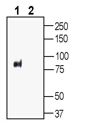
- Western blot analysis of rat kidney lysate: - 1. Anti-PepT2/SLC15A2 (extracellular) Antibody (#APT-002), (1:200). 2. Anti-PepT2/SLC15A2 (extracellular) Antibody , preincubated with PepT2/SLC15A2 (extracellular) Blocking Peptide (BLP-PT002).
- Submitted by
- Invitrogen Antibodies (provider)
- Main image
- Experimental details
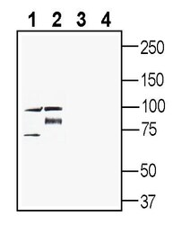
- Western blot analysis of rat brain membranes (lanes 1 and 3) and mouse brain membranes (lanes 2 and 4): - 1-2. Anti-PepT2/SLC15A2 (extracellular) Antibody (#APT-002), (1:500). 3-4. Anti-PepT2/SLC15A2 (extracellular) Antibody , preincubated with PepT2/SLC15A2 (extracellular) Blocking Peptide (BLP-PT002).
- Submitted by
- Invitrogen Antibodies (provider)
- Main image
- Experimental details
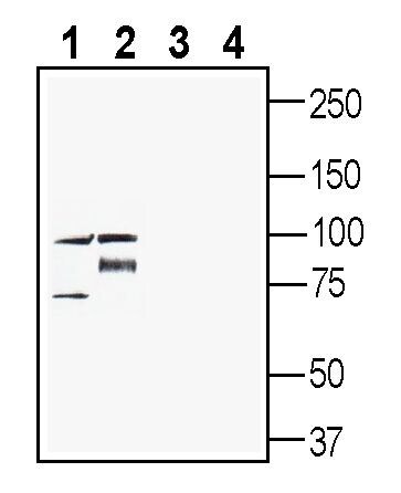
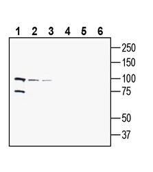
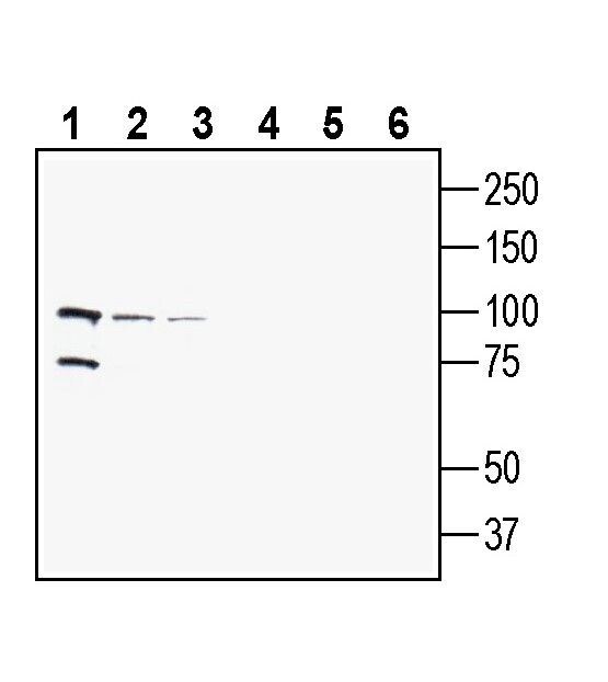
- Western blot analysis of human MCF-7 breast adenocarcinoma cell line lysate (lanes 1 and 4), human THP-1 monocytic leukemia cell line lysate (lanes 2 and 5) and human SH-SY5Y neuroblastoma cell line lysate (lanes 3 and 6): - 1-3. Anti-PepT2/SLC15A2 (extracellular) Antibody (#APT-002), (1:200). 4-6. Anti-PepT2/SLC15A2 (extracellular) Antibody , preincubated with PepT2/SLC15A2 (extracellular) Blocking Peptide (BLP-PT002).
Supportive validation
- Submitted by
- Invitrogen Antibodies (provider)
- Main image
- Experimental details
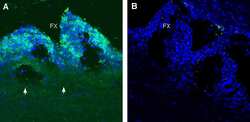
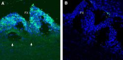
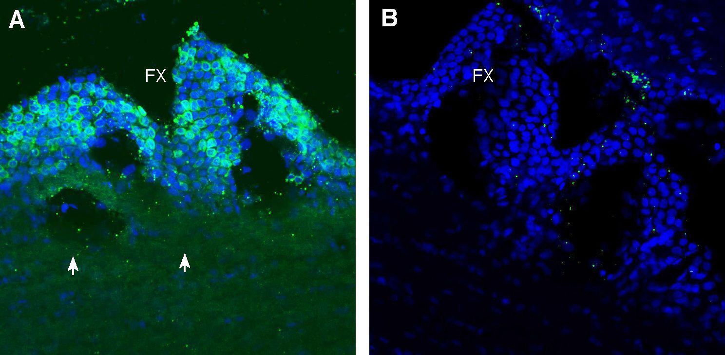
- Expression of PepT2/SLC15A2 in rat subfornical organ. Immunohistochemical staining of perfusion-fixed frozen rat brain sections with Anti-PepT2/SLC15A2 (extracellular) Antibody (#APT-002), (1:100), followed by goat Anti-rabbit-AlexaFluor-488. A. PepT2 immunoreactivity (green) appears in cells of the subfornical organ (arrows). B. Pre-incubation of the Antibody with PepT2/SLC15A2 (extracellular) Blocking Peptide (BLP-PT002), suppressed staining. Cell nuclei are stained with DAPI (blue). FX = fornix.
- Submitted by
- Invitrogen Antibodies (provider)
- Main image
- Experimental details
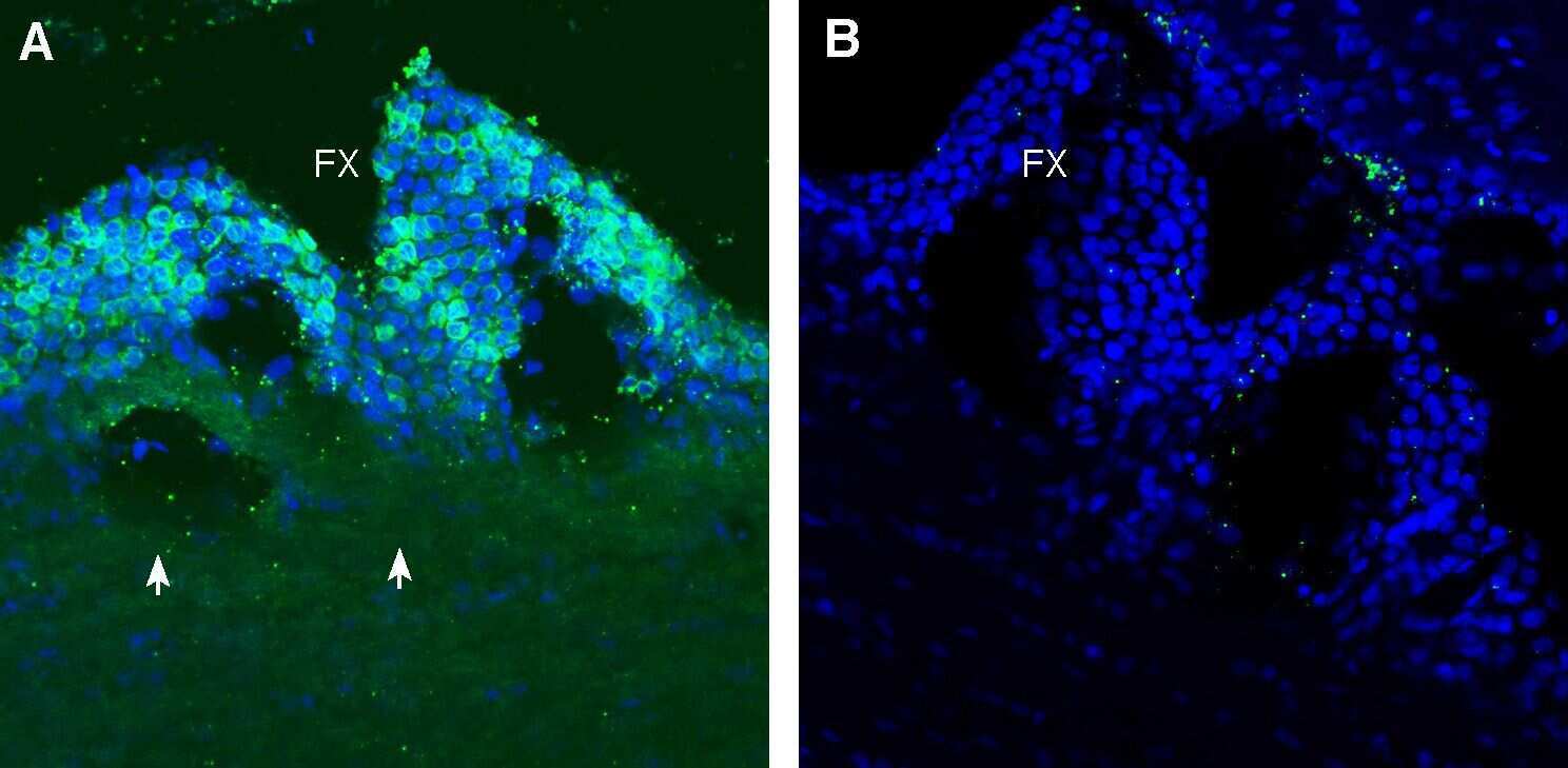
- Expression of PepT2/SLC15A2 in rat subfornical organ. Immunohistochemical staining of perfusion-fixed frozen rat brain sections with Anti-PepT2/SLC15A2 (extracellular) Antibody (#APT-002), (1:100), followed by goat Anti-rabbit-AlexaFluor-488. A. PepT2 immunoreactivity (green) appears in cells of the subfornical organ (arrows). B. Pre-incubation of the Antibody with PepT2/SLC15A2 (extracellular) Blocking Peptide (BLP-PT002), suppressed staining. Cell nuclei are stained with DAPI (blue). FX = fornix.
Supportive validation
- Submitted by
- Invitrogen Antibodies (provider)
- Main image
- Experimental details
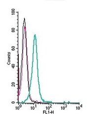
- Cell surface detection of PepT2/SLC15A2 by indirect flow cytometry in live intact human THP-1 monocytic leukemia cells: - (black line) cells. (red) Cells + goat- Anti-rabbit-FITC. (green) Cells + Anti-PepT2/SLC15A2 (extracellular) Antibody (#APT-002), (5μg) + goat- Anti-rabbit-FITC.
