Explore
 Validate
 Learn
Learn Western blot
 Immunocytochemistry
Antibody data
- Antibody Data
- Antigen structure
- References [55]
- Comments [0]
- Validations
- Immunocytochemistry [1]
Submit
Validation data
Reference
Comment
Report error
- Product number
- HPA021241 - Provider product page

- Provider
- Atlas Antibodies
- Proper citation
- Atlas Antibodies Cat#HPA021241, RRID:AB_1855299
- Product name
- Anti-PHGDH
- Antibody type
- Polyclonal
- Description
- Polyclonal Antibody against Human PHGDH, Gene description: phosphoglycerate dehydrogenase, Alternative Gene Names: PDG, PGDH, SERA, Validated applications: ICC, IHC, WB, Uniprot ID: O43175, Storage: Store at +4°C for short term storage. Long time storage is recommended at -20°C.
- Reactivity
- Human, Mouse, Rat
- Host
- Rabbit
- Conjugate
- Unconjugated
- Isotype
- IgG
- Vial size
- 100 µl
- Concentration
- 0.2 mg/ml
- Storage
- Store at +4°C for short term storage. Long time storage is recommended at -20°C.
- Handling
- The antibody solution should be gently mixed before use.
Submitted references Failure to repair damaged NAD(P)H blocks de novo serine synthesis in human cells
Central carbon metabolism exhibits unique characteristics during the handling of fungal patterns by monocyte-derived dendritic cells
ASCT2 is a major contributor to serine uptake in cancer cells
High expression of phosphoglycerate dehydrogenase predicts poor outcome in patients with high-grade serous ovarian cancer
Extracellular Vesicle Secretion by Leukemia Cells In Vivo Promotes CLL Progression by Hampering Antitumor T-cell Responses
Low glucose metabolite 3-phosphoglycerate switches PHGDH from serine synthesis to p53 activation to control cell fate
MicroRNA regulation of the serine synthesis pathway in endocrine-resistant breast cancer cells
p63 orchestrates serine and one carbon metabolism enzymes expression in head and neck cancer
PHGDH-mediated endothelial metabolism drives glioblastoma resistance to chimeric antigen receptor T cell immunotherapy.
An LKB1–mitochondria axis controls TH17 effector function
PHGDH heterogeneity potentiates cancer cell dissemination and metastasis
Lineage-specific silencing of PSAT1 induces serine auxotrophy and sensitivity to dietary serine starvation in luminal breast tumors
Glycolysis Dependency as a Hallmark of SF3B1-Mutated Cells
Mitochondria preserve an autarkic one-carbon cycle to confer growth-independent cancer cell migration and metastasis
Phosphoglycerate dehydrogenase positively regulates the proliferation of chicken muscle cells
Serine metabolism remodeling after platinum-based chemotherapy identifies vulnerabilities in a subgroup of resistant ovarian cancers.
In Vivo Evidence for Serine Biosynthesis-Defined Sensitivity of Lung Metastasis, but Not of Primary Breast Tumors, to mTORC1 Inhibition
Hepatic mTORC1 signaling activates ATF4 as part of its metabolic response to feeding and insulin
ATF3 promotes the serine synthesis pathway and tumor growth under dietary serine restriction
Metabolic compensation activates pro-survival mTORC1 signaling upon 3-phosphoglycerate dehydrogenase inhibition in osteosarcoma
Exercise-induced angiogenesis is dependent on metabolically primed ATF3/4+ endothelial cells
Enzymatic activation of pyruvate kinase increases cytosolic oxaloacetate to inhibit the Warburg effect
Serine biosynthesis defect due to haploinsufficiency of PHGDH causes retinal disease
Serine synthesis influences tamoxifen response in ER+ human breast carcinoma
One carbon metabolism in human lung cancer.
Serine synthesis pathway inhibition cooperates with dietary serine and glycine limitation for cancer therapy.
PHGDH supports liver ceramide synthesis and sustains lipid homeostasis
Inverse Data-Driven Modeling and Multiomics Analysis Reveals Phgdh as a Metabolic Checkpoint of Macrophage Polarization and Proliferation
Characterization of PHGDH expression in bladder cancer: potential targeting therapy with gemcitabine/cisplatin and the contribution of promoter DNA hypomethylation
Structure–Activity Relationships (SARs) of α-Ketothioamides as Inhibitors of Phosphoglycerate Dehydrogenase (PHGDH)
Macrophages induce malignant traits in mammary epithelium via IKKε/TBK1 kinases and the serine biosynthesis pathway
Limited Environmental Serine and Glycine Confer Brain Metastasis Sensitivity to PHGDH Inhibition
ILF3 is a substrate of SPOP for regulating serine biosynthesis in colorectal cancer
Increased Serine Synthesis Provides an Advantage for Tumors Arising in Tissues Where Serine Levels Are Limiting
Increased PHGDH expression promotes aberrant melanin accumulation
Hotspot SF3B1 mutations induce metabolic reprogramming and vulnerability to serine deprivation
Genome-wide CRISPR/Cas9 library screening identified PHGDH as a critical driver for Sorafenib resistance in HCC
Translational and HIF-1α-Dependent Metabolic Reprogramming Underpin Metabolic Plasticity and Responses to Kinase Inhibitors and Biguanides
PHGDH as a Key Enzyme for Serine Biosynthesis in HIF2α-Targeting Therapy for Renal Cell Carcinoma
Modulating the therapeutic response of tumours to dietary serine and glycine starvation
Hexokinases link DJ-1 to the PINK1/parkin pathway.
Validating and enabling phosphoglycerate dehydrogenase (PHGDH) as a target for fragment-based drug discovery in PHGDH-amplified breast cancer
Mitochondrial phosphoenolpyruvate carboxykinase (PEPCK-M) and serine biosynthetic pathway genes are co-ordinately increased during anabolic agent-induced skeletal muscle growth
A PHGDH inhibitor reveals coordination of serine synthesis and one-carbon unit fate
Identification of a small molecule inhibitor of 3-phosphoglycerate dehydrogenase to target serine biosynthesis in cancers
High level PHGDH expression in breast is predominantly associated with keratin 5‐positive cell lineage independently of malignancy
Reducing the serine availability complements the inhibition of the glutamine metabolism to block leukemia cell growth
NRF2 regulates serine biosynthesis in non–small cell lung cancer
Cells deficient in base-excision repair reveal cancer hallmarks originating from adjustments to genetic instability
An epitope tag alters phosphoglycerate dehydrogenase structure and impairs ability to support cell proliferation
p53 Protein-mediated Regulation of Phosphoglycerate Dehydrogenase (PHGDH) Is Crucial for the Apoptotic Response upon Serine Starvation
Mouse Genetics Suggests Cell-Context Dependency for Myc-Regulated Metabolic Enzymes during Tumorigenesis
Serine starvation induces stress and p53-dependent metabolic remodelling in cancer cells
Functional genomics reveal that the serine synthesis pathway is essential in breast cancer
Walvekar A, Warmoes M, Cheung D, Sikora T, Seyedkatouli N, Gomez-Giro G, Perrone S, Dengler L, Unger F, Santos B, Gavotto F, Dong X, Becker-Kettern J, Kwon Y, Jäger C, Schwamborn J, Van Bergen N, Christodoulou J, Linster C
Cellular & Molecular Biology Letters 2025;30(1)
Cellular & Molecular Biology Letters 2025;30(1)
Central carbon metabolism exhibits unique characteristics during the handling of fungal patterns by monocyte-derived dendritic cells
Alvarez Y, Mancebo C, Alonso S, Montero O, Fernández N, Sánchez Crespo M
Redox Biology 2024;73
Redox Biology 2024;73
ASCT2 is a major contributor to serine uptake in cancer cells
Conger K, Chidley C, Ozgurses M, Zhao H, Kim Y, Semina S, Burns P, Rawat V, Lietuvninkas L, Sheldon R, Ben-Sahra I, Frasor J, Sorger P, DeNicola G, Coloff J
Cell Reports 2024;43(8):114552
Cell Reports 2024;43(8):114552
High expression of phosphoglycerate dehydrogenase predicts poor outcome in patients with high-grade serous ovarian cancer
van Wagensveld L, Van Nyen T, Annibali D, Sonke G, Kruitwagen R, Amant F, Horlings H
The Oncologist 2024;29(9):e1231-e1234
The Oncologist 2024;29(9):e1231-e1234
Jasani N, Xu X, Posorske B, Kim Y, Vera O, Tsai K, DeNicola G, Karreth F
2024
2024
Extracellular Vesicle Secretion by Leukemia Cells In Vivo Promotes CLL Progression by Hampering Antitumor T-cell Responses
Gargiulo E, Viry E, Morande P, Largeot A, Gonder S, Xian F, Ioannou N, Benzarti M, Kleine Borgmann F, Mittelbronn M, Dittmar G, Nazarov P, Meiser J, Stamatopoulos B, Ramsay A, Moussay E, Paggetti J
Blood Cancer Discovery 2023;4(1):54-77
Blood Cancer Discovery 2023;4(1):54-77
Low glucose metabolite 3-phosphoglycerate switches PHGDH from serine synthesis to p53 activation to control cell fate
Wu Y, Zhang C, Xiong J, Cai D, Wang C, Wang Y, Liu Y, Wang Y, Li Y, Wu J, Wu J, Lan B, Wang X, Chen S, Cao X, Wei X, Hu H, Guo H, Yu Y, Ghafoor A, Xie C, Wu Y, Xu Z, Zhang C, Zhu M, Huang X, Sun X, Lin S, Piao H, Zhou J, Lin S
Cell Research 2023;33(11):835-850
Cell Research 2023;33(11):835-850
MicroRNA regulation of the serine synthesis pathway in endocrine-resistant breast cancer cells
Petri B, Piell K, Wilt A, Howser A, Winkler L, Whitworth M, Valdes B, Lehman N, Clem B, Klinge C
Endocrine-Related Cancer 2023;30(11)
Endocrine-Related Cancer 2023;30(11)
p63 orchestrates serine and one carbon metabolism enzymes expression in head and neck cancer
Cappello A, Tosetti G, Smirnov A, Ganini C, Yang X, Shi Y, Wang Y, Melino G, Bernassola F, Candi E
Biology Direct 2023;18(1)
Biology Direct 2023;18(1)
PHGDH-mediated endothelial metabolism drives glioblastoma resistance to chimeric antigen receptor T cell immunotherapy.
Zhang D, Li AM, Hu G, Huang M, Yang F, Zhang L, Wellen KE, Xu X, Conn CS, Zou W, Kahn M, Rhoades SD, Weljie AM, Fuchs SY, Amankulor N, Yoshor D, Ye J, Koumenis C, Gong Y, Fan Y
Cell metabolism 2023 Mar 7;35(3):517-534.e8
Cell metabolism 2023 Mar 7;35(3):517-534.e8
An LKB1–mitochondria axis controls TH17 effector function
Baixauli F, Piletic K, Puleston D, Villa M, Field C, Flachsmann L, Quintana A, Rana N, Edwards-Hicks J, Matsushita M, Stanczak M, Grzes K, Kabat A, Fabri M, Caputa G, Kelly B, Corrado M, Musa Y, Duda K, Mittler G, O’Sullivan D, Sesaki H, Jenuwein T, Buescher J, Pearce E, Sanin D, Pearce E
Nature 2022;610(7932):555-561
Nature 2022;610(7932):555-561
PHGDH heterogeneity potentiates cancer cell dissemination and metastasis
Rossi M, Altea-Manzano P, Demicco M, Doglioni G, Bornes L, Fukano M, Vandekeere A, Cuadros A, Fernández-García J, Riera-Domingo C, Jauset C, Planque M, Alkan H, Nittner D, Zuo D, Broadfield L, Parik S, Pane A, Rizzollo F, Rinaldi G, Zhang T, Teoh S, Aurora A, Karras P, Vermeire I, Broekaert D, Elsen J, Knott M, Orth M, Demeyer S, Eelen G, Dobrolecki L, Bassez A, Brussel T, Sotlar K, Lewis M, Bartsch H, Wuhrer M, Veelen P, Carmeliet P, Cools J, Morrison S, Marine J, Lambrechts D, Mazzone M, Hannon G, Lunt S, Grünewald T, Park M, Rheenen J, Fendt S
Nature 2022;605(7911):747-753
Nature 2022;605(7911):747-753
Lineage-specific silencing of PSAT1 induces serine auxotrophy and sensitivity to dietary serine starvation in luminal breast tumors
Choi B, Rawat V, Högström J, Burns P, Conger K, Ozgurses M, Patel J, Mehta T, Warren A, Selfors L, Muranen T, Coloff J
Cell Reports 2022;38(3):110278
Cell Reports 2022;38(3):110278
Glycolysis Dependency as a Hallmark of SF3B1-Mutated Cells
Vivet-Noguer R, Tarin M, Canbezdi C, Dayot S, Silva L, Houy A, Martineau S, Mieulet V, Gentric G, Loew D, Lombard B, Nemati F, Richon S, Guyonnet L, Servois V, Vagner S, Stern M, Roman-Roman S, Alsafadi S
Cancers 2022;14(9):2113
Cancers 2022;14(9):2113
Mitochondria preserve an autarkic one-carbon cycle to confer growth-independent cancer cell migration and metastasis
Kiweler N, Delbrouck C, Pozdeev V, Neises L, Soriano-Baguet L, Eiden K, Xian F, Benzarti M, Haase L, Koncina E, Schmoetten M, Jaeger C, Noman M, Vazquez A, Janji B, Dittmar G, Brenner D, Letellier E, Meiser J
Nature Communications 2022;13(1)
Nature Communications 2022;13(1)
Phosphoglycerate dehydrogenase positively regulates the proliferation of chicken muscle cells
Wang H, Hu M, Ding Z, Zhou X, Yang S, Shen Z, Yan F, Zhao A
Poultry Science 2022;101(5):101805
Poultry Science 2022;101(5):101805
Serine metabolism remodeling after platinum-based chemotherapy identifies vulnerabilities in a subgroup of resistant ovarian cancers.
Van Nyen T, Planque M, van Wagensveld L, Duarte JAG, Zaal EA, Talebi A, Rossi M, Körner PR, Rizzotto L, Moens S, De Wispelaere W, Baiden-Amissah REM, Sonke GS, Horlings HM, Eelen G, Berardi E, Swinnen JV, Berkers CR, Carmeliet P, Lambrechts D, Davidson B, Agami R, Fendt SM, Annibali D, Amant F
Nature communications 2022 Aug 5;13(1):4578
Nature communications 2022 Aug 5;13(1):4578
In Vivo Evidence for Serine Biosynthesis-Defined Sensitivity of Lung Metastasis, but Not of Primary Breast Tumors, to mTORC1 Inhibition
Rinaldi G, Pranzini E, Van Elsen J, Broekaert D, Funk C, Planque M, Doglioni G, Altea-Manzano P, Rossi M, Geldhof V, Teoh S, Ross C, Hunter K, Lunt S, Grünewald T, Fendt S
Molecular Cell 2021;81(2):386-397.e7
Molecular Cell 2021;81(2):386-397.e7
Hepatic mTORC1 signaling activates ATF4 as part of its metabolic response to feeding and insulin
Byles V, Cormerais Y, Kalafut K, Barrera V, Hughes Hallett J, Sui S, Asara J, Adams C, Hoxhaj G, Ben-Sahra I, Manning B
Molecular Metabolism 2021;53
Molecular Metabolism 2021;53
ATF3 promotes the serine synthesis pathway and tumor growth under dietary serine restriction
Li X, Gracilla D, Cai L, Zhang M, Yu X, Chen X, Zhang J, Long X, Ding H, Yan C
Cell Reports 2021;36(12):109706
Cell Reports 2021;36(12):109706
Metabolic compensation activates pro-survival mTORC1 signaling upon 3-phosphoglycerate dehydrogenase inhibition in osteosarcoma
Rathore R, Caldwell K, Schutt C, Brashears C, Prudner B, Ehrhardt W, Leung C, Lin H, Daw N, Beird H, Giles A, Wang W, Lazar A, Chrisinger J, Livingston J, Van Tine B
Cell Reports 2021;34(4):108678
Cell Reports 2021;34(4):108678
Exercise-induced angiogenesis is dependent on metabolically primed ATF3/4+ endothelial cells
Fan Z, Turiel G, Ardicoglu R, Ghobrial M, Masschelein E, Kocijan T, Zhang J, Tan G, Fitzgerald G, Gorski T, Alvarado-Diaz A, Gilardoni P, Adams C, Ghesquière B, De Bock K
Cell Metabolism 2021;33(9):1793-1807.e9
Cell Metabolism 2021;33(9):1793-1807.e9
Enzymatic activation of pyruvate kinase increases cytosolic oxaloacetate to inhibit the Warburg effect
Wiese E, Hitosugi S, Loa S, Sreedhar A, Andres-Beck L, Kurmi K, Pang Y, Karnitz L, Gonsalves W, Hitosugi T
Nature Metabolism 2021;3(7):954-968
Nature Metabolism 2021;3(7):954-968
Serine biosynthesis defect due to haploinsufficiency of PHGDH causes retinal disease
Eade K, Gantner M, Hostyk J, Nagasaki T, Giles S, Fallon R, Harkins-Perry S, Baldini M, Lim E, Scheppke L, Dorrell M, Cai C, Baugh E, Wolock C, Wallace M, Berlow R, Goldstein D, Metallo C, Friedlander M, Allikmets R
Nature Metabolism 2021;3(3):366-377
Nature Metabolism 2021;3(3):366-377
Serine synthesis influences tamoxifen response in ER+ human breast carcinoma
Metcalf S, Petri B, Kruer T, Green B, Dougherty S, Wittliff J, Klinge C, Clem B
Endocrine-Related Cancer 2021;28(1):27-37
Endocrine-Related Cancer 2021;28(1):27-37
One carbon metabolism in human lung cancer.
Yao S, Peng L, Elakad O, Küffer S, Hinterthaner M, Danner BC, von Hammerstein-Equord A, Ströbel P, Bohnenberger H
Translational lung cancer research 2021 Jun;10(6):2523-2538
Translational lung cancer research 2021 Jun;10(6):2523-2538
Serine synthesis pathway inhibition cooperates with dietary serine and glycine limitation for cancer therapy.
Tajan M, Hennequart M, Cheung EC, Zani F, Hock AK, Legrave N, Maddocks ODK, Ridgway RA, Athineos D, Suárez-Bonnet A, Ludwig RL, Novellasdemunt L, Angelis N, Li VSW, Vlachogiannis G, Valeri N, Mainolfi N, Suri V, Friedman A, Manfredi M, Blyth K, Sansom OJ, Vousden KH
Nature communications 2021 Jan 14;12(1):366
Nature communications 2021 Jan 14;12(1):366
PHGDH supports liver ceramide synthesis and sustains lipid homeostasis
Kang Y, Falzone A, Liu M, González-Sánchez P, Choi B, Coloff J, Saller J, Karreth F, DeNicola G
Cancer & Metabolism 2020;8(1)
Cancer & Metabolism 2020;8(1)
Inverse Data-Driven Modeling and Multiomics Analysis Reveals Phgdh as a Metabolic Checkpoint of Macrophage Polarization and Proliferation
Wilson J, Nägele T, Linke M, Demel F, Fritsch S, Mayr H, Cai Z, Katholnig K, Sun X, Fragner L, Miller A, Haschemi A, Popa A, Bergthaler A, Hengstschläger M, Weichhart T, Weckwerth W
Cell Reports 2020;30(5):1542-1552.e7
Cell Reports 2020;30(5):1542-1552.e7
Characterization of PHGDH expression in bladder cancer: potential targeting therapy with gemcitabine/cisplatin and the contribution of promoter DNA hypomethylation
Yoshino H, Enokida H, Osako Y, Nohata N, Yonemori M, Sugita S, Kuroshima K, Tsuruda M, Tatarano S, Nakagawa M
Molecular Oncology 2020;14(9):2190-2202
Molecular Oncology 2020;14(9):2190-2202
Structure–Activity Relationships (SARs) of α-Ketothioamides as Inhibitors of Phosphoglycerate Dehydrogenase (PHGDH)
Spillier Q, Ravez S, Unterlass J, Corbet C, Degavre C, Feron O, Frédérick R
Pharmaceuticals 2020;13(2):20
Pharmaceuticals 2020;13(2):20
Macrophages induce malignant traits in mammary epithelium via IKKε/TBK1 kinases and the serine biosynthesis pathway
Wilcz‐Villega E, Carter E, Ironside A, Xu R, Mataloni I, Holdsworth J, Jones W, Moreno Béjar R, Uhlik L, Bentham R, Godinho S, Dalli J, Grose R, Szabadkai G, Jones L, Hodivala‐Dilke K, Bianchi K
EMBO Molecular Medicine 2020;12(2)
EMBO Molecular Medicine 2020;12(2)
Limited Environmental Serine and Glycine Confer Brain Metastasis Sensitivity to PHGDH Inhibition
Ngo B, Kim E, Osorio-Vasquez V, Doll S, Bustraan S, Liang R, Luengo A, Davidson S, Ali A, Ferraro G, Fischer G, Eskandari R, Kang D, Ni J, Plasger A, Rajasekhar V, Kastenhuber E, Bacha S, Sriram R, Stein B, Bakhoum S, Snuderl M, Cotzia P, Healey J, Mainolfi N, Suri V, Friedman A, Manfredi M, Sabatini D, Jones D, Yu M, Zhao J, Jain R, Keshari K, Davies M, Vander Heiden M, Hernando E, Mann M, Cantley L, Pacold M
Cancer Discovery 2020;10(9):1352-1373
Cancer Discovery 2020;10(9):1352-1373
ILF3 is a substrate of SPOP for regulating serine biosynthesis in colorectal cancer
Li K, Wu J, Qin B, Fan Z, Tang Q, Lu W, Zhang H, Xing F, Meng M, Zou S, Wei W, Chen H, Cai J, Wang H, Zhang H, Cai J, Fang L, Bian X, Chen C, Lan P, Ghesquière B, Fang L, Lee M
Cell Research 2019;30(2):163-178
Cell Research 2019;30(2):163-178
Increased Serine Synthesis Provides an Advantage for Tumors Arising in Tissues Where Serine Levels Are Limiting
Sullivan M, Mattaini K, Dennstedt E, Nguyen A, Sivanand S, Reilly M, Meeth K, Muir A, Darnell A, Bosenberg M, Lewis C, Vander Heiden M
Cell Metabolism 2019;29(6):1410-1421.e4
Cell Metabolism 2019;29(6):1410-1421.e4
Increased PHGDH expression promotes aberrant melanin accumulation
Mattaini K, Sullivan M, Lau A, Fiske B, Bronson R, Vander Heiden M
BMC Cancer 2019;19(1)
BMC Cancer 2019;19(1)
Hotspot SF3B1 mutations induce metabolic reprogramming and vulnerability to serine deprivation
Dalton W, Helmenstine E, Walsh N, Gondek L, Kelkar D, Read A, Natrajan R, Christenson E, Roman B, Das S, Zhao L, Leone R, Shinn D, Groginski T, Madugundu A, Patil A, Zabransky D, Medford A, Lee J, Cole A, Rosen M, Thakar M, Ambinder A, Donaldson J, DeZern A, Cravero K, Chu D, Madero-Marroquin R, Pandey A, Hurley P, Lauring J, Park B
Journal of Clinical Investigation 2019;129(11):4708-4723
Journal of Clinical Investigation 2019;129(11):4708-4723
Genome-wide CRISPR/Cas9 library screening identified PHGDH as a critical driver for Sorafenib resistance in HCC
Wei L, Lee D, Law C, Zhang M, Shen J, Chin D, Zhang A, Tsang F, Wong C, Ng I, Wong C, Wong C
Nature Communications 2019;10(1)
Nature Communications 2019;10(1)
Translational and HIF-1α-Dependent Metabolic Reprogramming Underpin Metabolic Plasticity and Responses to Kinase Inhibitors and Biguanides
Hulea L, Gravel S, Morita M, Cargnello M, Uchenunu O, Im Y, Lehuédé C, Ma E, Leibovitch M, McLaughlan S, Blouin M, Parisotto M, Papavasiliou V, Lavoie C, Larsson O, Ohh M, Ferreira T, Greenwood C, Bridon G, Avizonis D, Ferbeyre G, Siegel P, Jones R, Muller W, Ursini-Siegel J, St-Pierre J, Pollak M, Topisirovic I
Cell Metabolism 2018;28(6):817-832.e8
Cell Metabolism 2018;28(6):817-832.e8
PHGDH as a Key Enzyme for Serine Biosynthesis in HIF2α-Targeting Therapy for Renal Cell Carcinoma
Yoshino H, Nohata N, Miyamoto K, Yonemori M, Sakaguchi T, Sugita S, Itesako T, Kofuji S, Nakagawa M, Dahiya R, Enokida H
Cancer Research 2017;77(22):6321-6329
Cancer Research 2017;77(22):6321-6329
Modulating the therapeutic response of tumours to dietary serine and glycine starvation
Maddocks O, Athineos D, Cheung E, Lee P, Zhang T, van den Broek N, Mackay G, Labuschagne C, Gay D, Kruiswijk F, Blagih J, Vincent D, Campbell K, Ceteci F, Sansom O, Blyth K, Vousden K
Nature 2017;544(7650):372-376
Nature 2017;544(7650):372-376
Hexokinases link DJ-1 to the PINK1/parkin pathway.
Hauser DN, Mamais A, Conti MM, Primiani CT, Kumaran R, Dillman AA, Langston RG, Beilina A, Garcia JH, Diaz-Ruiz A, Bernier M, Fiesel FC, Hou X, Springer W, Li Y, de Cabo R, Cookson MR
Molecular neurodegeneration 2017 Sep 29;12(1):70
Molecular neurodegeneration 2017 Sep 29;12(1):70
Validating and enabling phosphoglycerate dehydrogenase (PHGDH) as a target for fragment-based drug discovery in PHGDH-amplified breast cancer
Unterlass J, Baslé A, Blackburn T, Tucker J, Cano C, Noble M, Curtin N
Oncotarget 2016;9(17):13139-13153
Oncotarget 2016;9(17):13139-13153
Mitochondrial phosphoenolpyruvate carboxykinase (PEPCK-M) and serine biosynthetic pathway genes are co-ordinately increased during anabolic agent-induced skeletal muscle growth
Brown D, Williams H, Ryan K, Wilson T, Daniel Z, Mareko M, Emes R, Harris D, Jones S, Wattis J, Dryden I, Hodgman T, Brameld J, Parr T
Scientific Reports 2016;6(1)
Scientific Reports 2016;6(1)
A PHGDH inhibitor reveals coordination of serine synthesis and one-carbon unit fate
Pacold M, Brimacombe K, Chan S, Rohde J, Lewis C, Swier L, Possemato R, Chen W, Sullivan L, Fiske B, Cho S, Freinkman E, Birsoy K, Abu-Remaileh M, Shaul Y, Liu C, Zhou M, Koh M, Chung H, Davidson S, Luengo A, Wang A, Xu X, Yasgar A, Liu L, Rai G, Westover K, Vander Heiden M, Shen M, Gray N, Boxer M, Sabatini D
Nature Chemical Biology 2016;12(6):452-458
Nature Chemical Biology 2016;12(6):452-458
Identification of a small molecule inhibitor of 3-phosphoglycerate dehydrogenase to target serine biosynthesis in cancers
Mullarky E, Lucki N, Beheshti Zavareh R, Anglin J, Gomes A, Nicolay B, Wong J, Christen S, Takahashi H, Singh P, Blenis J, Warren J, Fendt S, Asara J, DeNicola G, Lyssiotis C, Lairson L, Cantley L
Proceedings of the National Academy of Sciences 2016;113(7):1778-1783
Proceedings of the National Academy of Sciences 2016;113(7):1778-1783
High level PHGDH expression in breast is predominantly associated with keratin 5‐positive cell lineage independently of malignancy
Gromova I, Gromov P, Honma N, Kumar S, Rimm D, Talman M, Wielenga V, Moreira J
Molecular Oncology 2015;9(8):1636-1654
Molecular Oncology 2015;9(8):1636-1654
Reducing the serine availability complements the inhibition of the glutamine metabolism to block leukemia cell growth
Polet F, Corbet C, Pinto A, Rubio L, Martherus R, Bol V, Drozak X, Grégoire V, Riant O, Feron O
Oncotarget 2015;7(2):1765-1776
Oncotarget 2015;7(2):1765-1776
NRF2 regulates serine biosynthesis in non–small cell lung cancer
DeNicola G, Chen P, Mullarky E, Sudderth J, Hu Z, Wu D, Tang H, Xie Y, Asara J, Huffman K, Wistuba I, Minna J, DeBerardinis R, Cantley L
Nature Genetics 2015;47(12):1475-1481
Nature Genetics 2015;47(12):1475-1481
Cells deficient in base-excision repair reveal cancer hallmarks originating from adjustments to genetic instability
Markkanen E, Fischer R, Ledentcova M, Kessler B, Dianov G
Nucleic Acids Research 2015;43(7):3667-3679
Nucleic Acids Research 2015;43(7):3667-3679
An epitope tag alters phosphoglycerate dehydrogenase structure and impairs ability to support cell proliferation
Mattaini K, Brignole E, Kini M, Davidson S, Fiske B, Drennan C, Vander Heiden M
Cancer & Metabolism 2015;3(1)
Cancer & Metabolism 2015;3(1)
p53 Protein-mediated Regulation of Phosphoglycerate Dehydrogenase (PHGDH) Is Crucial for the Apoptotic Response upon Serine Starvation
Ou Y, Wang S, Jiang L, Zheng B, Gu W
Journal of Biological Chemistry 2015;290(1):457-466
Journal of Biological Chemistry 2015;290(1):457-466
Mouse Genetics Suggests Cell-Context Dependency for Myc-Regulated Metabolic Enzymes during Tumorigenesis
Clurman B, Nilsson L, Plym Forshell T, Rimpi S, Kreutzer C, Pretsch W, Bornkamm G, Nilsson J
PLoS Genetics 2012;8(3):e1002573
PLoS Genetics 2012;8(3):e1002573
Serine starvation induces stress and p53-dependent metabolic remodelling in cancer cells
Maddocks O, Berkers C, Mason S, Zheng L, Blyth K, Gottlieb E, Vousden K
Nature 2012;493(7433):542-546
Nature 2012;493(7433):542-546
Functional genomics reveal that the serine synthesis pathway is essential in breast cancer
Possemato R, Marks K, Shaul Y, Pacold M, Kim D, Birsoy K, Sethumadhavan S, Woo H, Jang H, Jha A, Chen W, Barrett F, Stransky N, Tsun Z, Cowley G, Barretina J, Kalaany N, Hsu P, Ottina K, Chan A, Yuan B, Garraway L, Root D, Mino-Kenudson M, Brachtel E, Driggers E, Sabatini D
Nature 2011;476(7360):346-350
Nature 2011;476(7360):346-350
No comments: Submit comment
Supportive validation
- Submitted by
- Atlas Antibodies (provider)
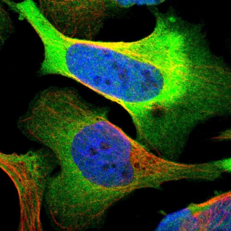
- Main image

- Experimental details
- Immunofluorescent staining of human cell line U-2 OS shows localization to plasma membrane & cytosol.
- Sample type
- Human