 Explore
 Validate
 Learn
Learn Western blot
 Immunocytochemistry
Antibody data
- Antibody Data
- Antigen structure
- References [2]
- Comments [0]
- Validations
- Immunocytochemistry [3]
- Immunoprecipitation [1]
- Immunohistochemistry [4]
- Other assay [2]
Submit
Validation data
Reference
Comment
Report error
- Product number
- PA5-28169 - Provider product page

- Provider
- Invitrogen Antibodies
- Product name
- MTHFD2 Polyclonal Antibody
- Antibody type
- Polyclonal
- Antigen
- Recombinant protein fragment
- Description
- Recommended positive controls: 293T, A431, H1299, HeLa, HepG2, Molt-4, Raji, SK-N-DZ, SK-N-SH. Predicted reactivity: Mouse (95%), Rat (93%), Zebrafish (86%), Xenopus laevis (90%), Chicken (92%), Bovine (95%). Store product as a concentrated solution. Centrifuge briefly prior to opening the vial.
- Reactivity
- Human
- Host
- Rabbit
- Isotype
- IgG
- Vial size
- 100 μL
- Concentration
- 0.13 mg/mL
- Storage
- Store at 4°C short term. For long term storage, store at -20°C, avoiding freeze/thaw cycles.
Submitted references MTHFD2 facilitates breast cancer cell proliferation via the AKT signaling pathway.
Cardiac metabolic pathways affected in the mouse model of barth syndrome.
Huang J, Qin Y, Lin C, Huang X, Zhang F
Experimental and therapeutic medicine 2021 Jul;22(1):703
Experimental and therapeutic medicine 2021 Jul;22(1):703
Cardiac metabolic pathways affected in the mouse model of barth syndrome.
Huang Y, Powers C, Madala SK, Greis KD, Haffey WD, Towbin JA, Purevjav E, Javadov S, Strauss AW, Khuchua Z
PloS one 2015;10(6):e0128561
PloS one 2015;10(6):e0128561
No comments: Submit comment
Supportive validation
- Submitted by
- Invitrogen Antibodies (provider)
- Main image

- Experimental details
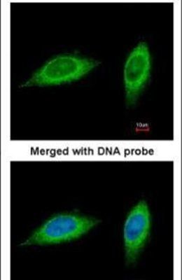
- Immunofluorescent analysis of MTHFD2 in paraformaldehyde-fixed HeLa cells using a MTHFD2 polyclonal antibody (Product # PA5-28169) at a 1:200 dilution.
- Submitted by
- Invitrogen Antibodies (provider)
- Main image
- Experimental details
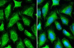
- MTHFD2 Polyclonal Antibody detects MTHFD2 protein at mitochondria by immunofluorescent analysis. Sample: HeLa cells were fixed in 4% paraformaldehyde at RT for 15 min. Green: MTHFD2 stained by MTHFD2 Polyclonal Antibody (Product # PA5-28169) diluted at 1:500. Blue: Fluoroshield with DAPI .

- Submitted by
- Invitrogen Antibodies (provider)
- Main image

- Experimental details
- MTHFD2 Polyclonal Antibody detects MTHFD2 protein at mitochondria by immunofluorescent analysis. Sample: HeLa cells were fixed in 4% paraformaldehyde at RT for 15 min. Green: MTHFD2 stained by MTHFD2 Polyclonal Antibody (Product # PA5-28169) diluted at 1:500. Blue: Fluoroshield with DAPI .

Supportive validation
- Submitted by
- Invitrogen Antibodies (provider)
- Main image
- Experimental details
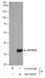
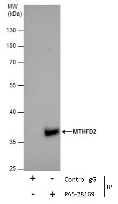
- Immunoprecipitation of MTHFD2 was performed in 293T whole cell extracts using 5 µg of MTHFD2 Polyclonal Antibody (Product # PA5-28169). Samples were transferred to a membrane and probed with MTHFD2 Polyclonal Antibody as a primary antibody and an HRP-conjugated anti-Rabbit IgG was used as a secondary antibody.
Supportive validation
- Submitted by
- Invitrogen Antibodies (provider)
- Main image
- Experimental details
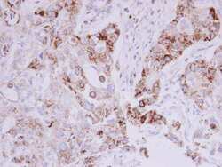
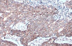
- Immunohistochemistry (Paraffin) analysis of MTHFD2 was performed in paraffin-embedded human breast carcinoma tissue using MTHFD2 Polyclonal Antibody (Product # PA5-28169) at a dilution of 1:500.
- Submitted by
- Invitrogen Antibodies (provider)
- Main image
- Experimental details
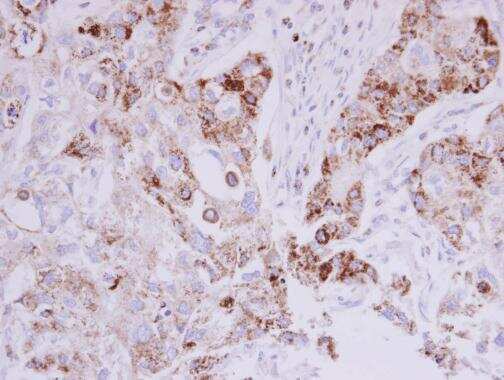
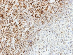
- Immunohistochemistry (Paraffin) analysis of MTHFD2 was performed in paraffin-embedded human MCF7 xenograft tissue using MTHFD2 Polyclonal Antibody (Product # PA5-28169) at a dilution of 1:500.
- Submitted by
- Invitrogen Antibodies (provider)
- Main image
- Experimental details
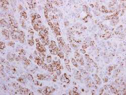
- MTHFD2 Polyclonal Antibody detects MTHFD2 protein at mitochondria by immunohistochemical analysis. Sample: Paraffin-embedded human colon cancer. MTHFD2 stained by MTHFD2 Polyclonal Antibody (Product # PA5-28169) diluted at 1:500. Antigen Retrieval: Citrate buffer, pH 6.0, 15 min.

- Submitted by
- Invitrogen Antibodies (provider)
- Main image
- Experimental details
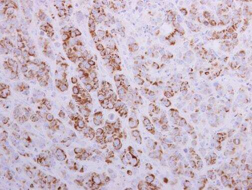
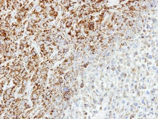
- Immunohistochemical analysis of paraffin-embedded SAS xenograft, using MTHFD2 (Product # PA5-28169) antibody at 1:500 dilution. Antigen Retrieval: EDTA based buffer, pH 8.0, 15 min.
Supportive validation
- Submitted by
- Invitrogen Antibodies (provider)
- Main image

- Experimental details
- Immunoprecipitation of MTHFD2 was performed in 293T whole cell extracts using 5 µg of MTHFD2 Polyclonal Antibody (Product # PA5-28169). Samples were transferred to a membrane and probed with MTHFD2 Polyclonal Antibody as a primary antibody and an HRP-conjugated anti-Rabbit IgG was used as a secondary antibody.

- Submitted by
- Invitrogen Antibodies (provider)
- Main image
- Experimental details
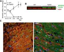
- Fig 5 Altered Mthfd2 expression in adult TazKD heart. (A) Quantification of Mthfd2 mRNA levels in hearts of WT and TazKD mice during development. (B) Immunoblots of total protein extracts from 3 month-old WT and TazKD hearts with anti-MTHFD2 antibodies (green). Antibodies specific to mitochondrial malate dehydrogenase (MDH) were used as a loading control (red). (C) Immunofluorescent staining of MTHFD2 in LV sections of 3 month-old WT and TazKD mice revealing localization of MTHFD2 protein (green) in intercalated disks (arrowheads) and myofilaments ( MF ). Mitochondria were visualized with antibodies specific to complex I subunit NDUFA9 (red). Nuclei were stained with DAPI (blue).
