Explore
 Validate
 Learn
Learn Western blot
 Immunoprecipitation
Antibody data
- Antibody Data
- Antigen structure
- References [0]
- Comments [0]
- Validations
- Western blot [5]
- Immunohistochemistry [2]
- Other assay [1]
Submit
Validation data
Reference
Comment
Report error
- Product number
- A300-864A - Provider product page

- Provider
- Invitrogen Antibodies
- Product name
- RBM9 Polyclonal Antibody
- Antibody type
- Polyclonal
- Antigen
- Other
- Reactivity
- Human, Mouse, Rat
- Host
- Rabbit
- Isotype
- IgG
- Vial size
- 100 µL
- Concentration
- 1 mg/mL
- Storage
- 4° C
No comments: Submit comment
Supportive validation
- Submitted by
- Invitrogen Antibodies (provider)
- Main image
- Experimental details
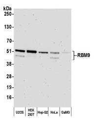
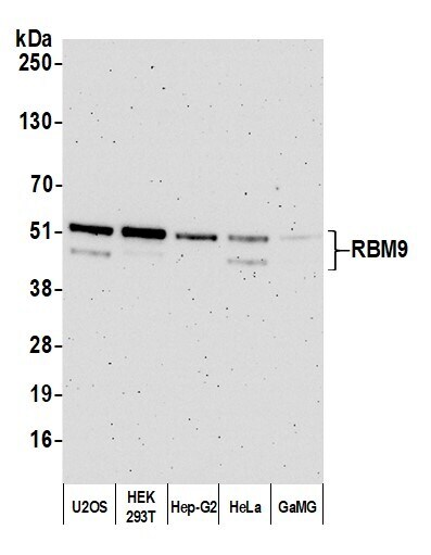
- Detection of human RBM9 by western blot. Samples: Whole cell lysate (10 µg) from U2OS, HEK293T, Hep-G2, HeLa, and GaMG cells prepared using NETN lysis buffer. Antibody: Affinity purified rabbit anti-RBM9 antibody (Product # A300-864A lot 4) used for WB at 0.1 µg/mL. Detection: Chemiluminescence with an exposure time of 3 minutes.
- Submitted by
- Invitrogen Antibodies (provider)
- Main image
- Experimental details
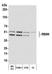
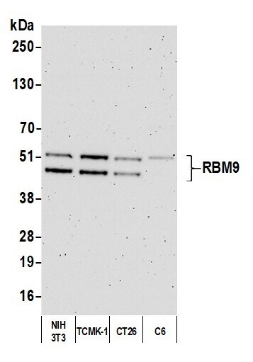
- Detection of human RBM9 by western blot. Samples: Whole cell lysate (10 µg) from NIH 3T3, TCMK-1, CT26, and C6 cells prepared using NETN lysis buffer. Antibody: Affinity purified rabbit anti-RBM9 antibody (Product # A300-864A; lot A300-864A-4) used for WB at 0.1 µg/mL. Detection: Chemiluminescence with an exposure time of 3 minutes.
- Submitted by
- Invitrogen Antibodies (provider)
- Main image
- Experimental details
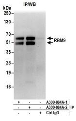
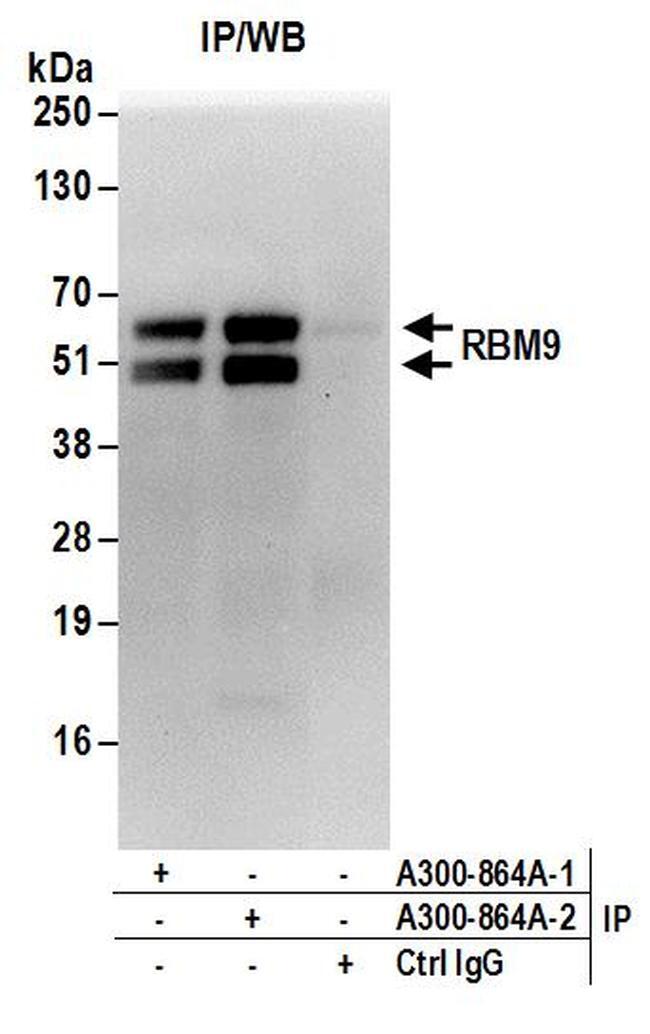
- Detection of human RBM9 by western blot of immunoprecipitates. Samples: Whole cell lysate (1 mg for IP; 20% of IP loaded) from HeLa cells. Antibodies: Affinity purified rabbit anti-RBM9 antibody A300-864A (lot A300-864A-2) used for IP at 6 µg/mg lysate. RBM9 was also immunoprecipitated by a previous lot (lot A300-864A-1) of this antibody. For blotting immunoprecipitated RBM9, A300-864A-2 was used at 1 µg/ml. Detection: Chemiluminescence with an exposure time of 30 seconds.
- Submitted by
- Invitrogen Antibodies (provider)
- Main image
- Experimental details
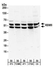
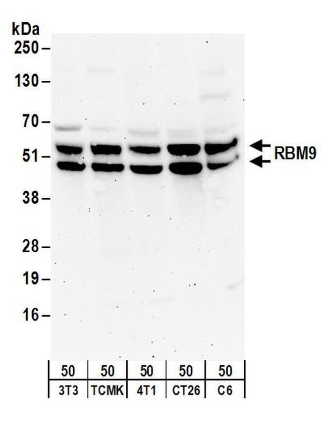
- Detection of mouse and rat RBM9 by western blot. Samples: Whole cell lysate (50 µg) from NIH3T3, TCMK-1, 4T1, CT26.WT, and rat C6 cells. Antibodies: Affinity purified rabbit anti-RBM9 antibody A300-864A (lot A300-864A-2) used for WB at 0.5 µg/ml. Detection: Chemiluminescence with an exposure time of 3 minutes.
- Submitted by
- Invitrogen Antibodies (provider)
- Main image
- Experimental details
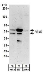
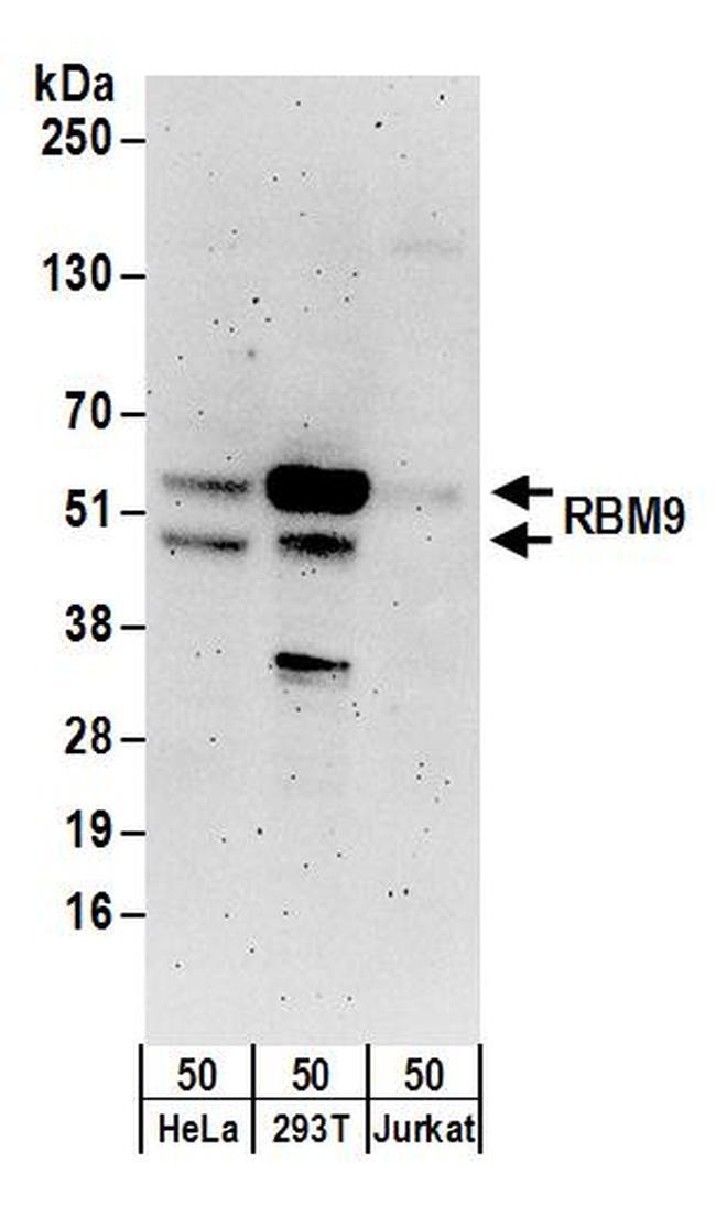
- Detection of human RBM9 by western blot. Samples: Whole cell lysate (50 µg) from HeLa, 293T, and Jurkat cells. Antibodies: Affinity purified rabbit anti-RBM9 antibody A300-864A (lot A300-864A-2) used for WB at 0.1 µg/ml. Detection: Chemiluminescence with an exposure time of 3 minutes.
Supportive validation
- Submitted by
- Invitrogen Antibodies (provider)
- Main image

- Experimental details
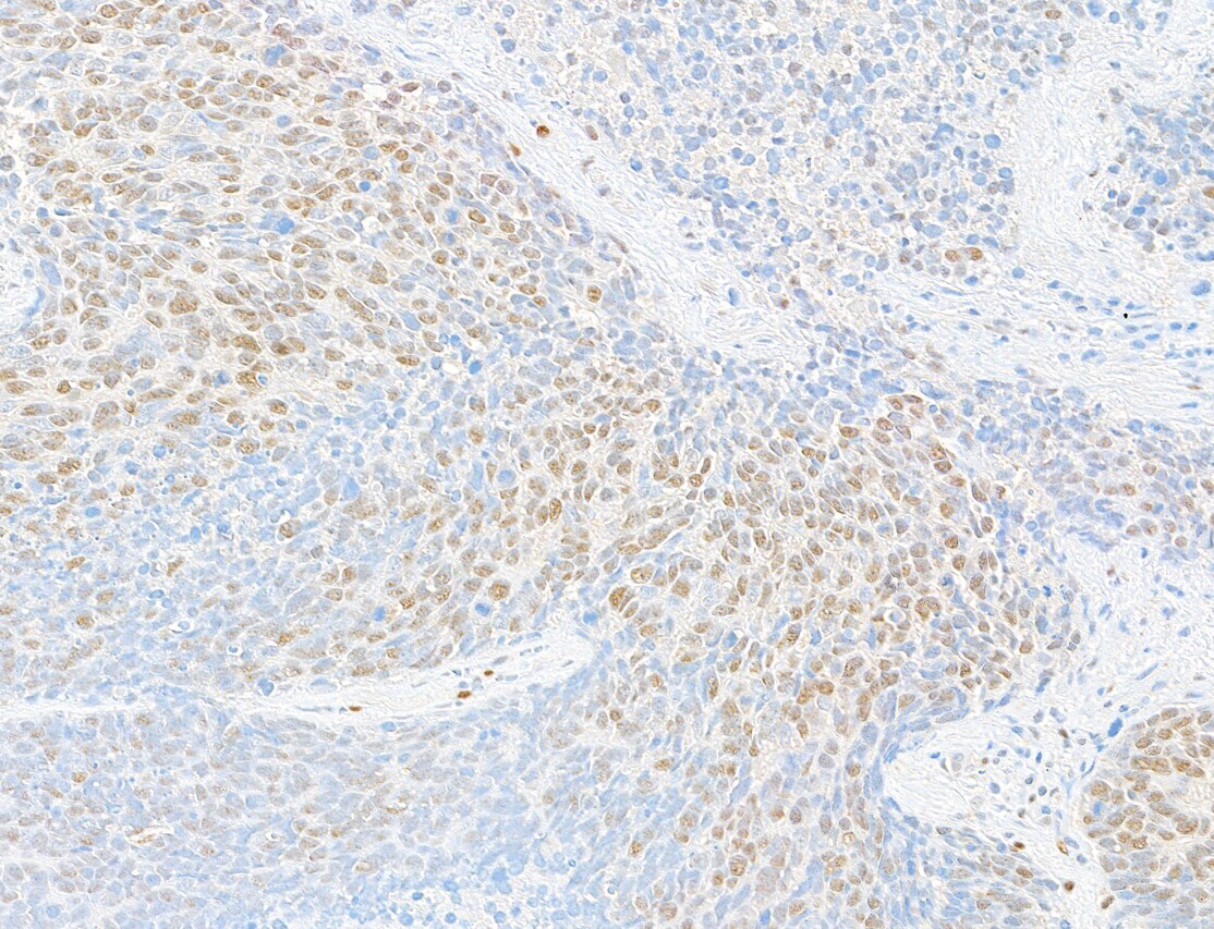
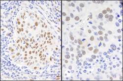
- Detection of human RBM9 by immunohistochemistry. Sample: FFPE section of human lung carcinoma. Antibody: Affinity purified rabbit anti-RBM9 (Product # A300-864A; lot 4) used at a dilution of 1:5,000 (0.2 µg/mL). Detection: DAB.
- Submitted by
- Invitrogen Antibodies (provider)
- Main image
- Experimental details
- Detection of human and mouse RBM9 by immunohistochemistry. Sample: FFPE sections of human lung carcinoma (left) and mouse renal cell carcinoma (right). Antibody: Affinity purified rabbit anti-RBM9 (Cat. No. A300-864A Lot2) used at a dilution of 1:1,000 (1µg/ml). Detection: DAB.

Supportive validation
- Submitted by
- Invitrogen Antibodies (provider)
- Main image
- Experimental details
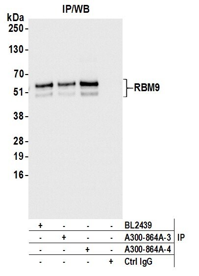
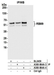
- Detection of human RBM9 by western blot of immunoprecipitates. Samples: Whole cell lysate (1.0 mg per IP reaction; 20% of IP loaded) from HEK293T cells prepared using NETN lysis buffer. Antibodies: Affinity purified rabbit anti-RBM9 antibody (Product # A300-864A lot 4) used for IP at 6 µg per reaction. RBM9 was also immunoprecipitated by a previous lot of this antibody (A300-864A lot 3) and a second antibody to a different epitope of RBM9 (BL2440). For blotting immunoprecipitated RBM9 (Product # A300-864A) was used at 0.1 µg/mL. Detection: Chemiluminescence with an exposure time of 1 second.