Explore
 Validate
 Learn
Learn14-9321-80
antibody from Invitrogen Antibodies
Targeting: F11R
CD321, JAM-1, JAM-A, JAM1, JAMA, JCAM, PAM-1
 Western blot
 Immunoprecipitation
Antibody data
- Antibody Data
- Antigen structure
- References [3]
- Comments [0]
- Validations
- Western blot [1]
- Immunocytochemistry [1]
- Flow cytometry [1]
Submit
Validation data
Reference
Comment
Report error
- Product number
- 14-9321-80 - Provider product page

- Provider
- Invitrogen Antibodies
- Product name
- CD321 (F11R) Monoclonal Antibody (WK9), eBioscience™
- Antibody type
- Monoclonal
- Antigen
- Other
- Description
- Description: CD321 (F11R, JAM-1, JAM-A) is a cell-surface adhesion molecule belonging to the Ig superfamily of receptors. CD321 is a 32-41 kD glycoprotein, possessing two extracellular Ig-like domains, a transmembrane region, and a short cytosolic tail. The protein assembles at the membrane as a homodimer, and can associate laterally with other membrane proteins including integrins. CD321 is expressed on a variety of cell types, including leukocytes, platelets, erythrocytes, epithelial and endothelial cells, and plays a role in mediating cell-cell contact. CD321 can be found in close proximity to several components of epithelial tight junctions, including ZO-1 and occludin. CD321 is also involved in reovirus entry into cells. Applications Reported: This WK9 antibody has been reported for use in flow cytometric analysis and immunoprecipitation. The WK9 antibody has also been found useful for immunoblotting, recognizing a protein of approximately 38 kD under reducing and non-reducing conditions. Applications Tested: This WK9 antibody has been tested by flow cytometric analysis of normal human peripheral blood cells. This can be used at less than or equal to 0.25 µg per test. A test is defined as the amount (µg) of antibody that will stain a cell sample in a final volume of 100 µL. Cell number should be determined empirically but can range from 10^5 to 10^8 cells/test. It is recommended that the antibody be carefully titrated for optimal performance in the assay of interest. Purity: Greater than 90%, as determined by SDS-PAGE. Aggregation: Less than 10%, as determined by HPLC. Filtration: 0.2 µm post-manufacturing filtered.
- Reactivity
- Human
- Host
- Mouse
- Isotype
- IgG
- Antibody clone number
- WK9
- Vial size
- 25 µg
- Concentration
- 0.5 mg/mL
- Storage
- 4° C
Submitted references Signaling through JAM-1 and alphavbeta3 is required for the angiogenic action of bFGF: dissociation of the JAM-1 and alphavbeta3 complex.
JAM-1 is a ligand of the beta(2) integrin LFA-1 involved in transendothelial migration of leukocytes.
Cloning of the human platelet F11 receptor: a cell adhesion molecule member of the immunoglobulin superfamily involved in platelet aggregation.
Naik MU, Mousa SA, Parkos CA, Naik UP
Blood 2003 Sep 15;102(6):2108-14
Blood 2003 Sep 15;102(6):2108-14
JAM-1 is a ligand of the beta(2) integrin LFA-1 involved in transendothelial migration of leukocytes.
Ostermann G, Weber KS, Zernecke A, Schröder A, Weber C
Nature immunology 2002 Feb;3(2):151-8
Nature immunology 2002 Feb;3(2):151-8
Cloning of the human platelet F11 receptor: a cell adhesion molecule member of the immunoglobulin superfamily involved in platelet aggregation.
Sobocka MB, Sobocki T, Banerjee P, Weiss C, Rushbrook JI, Norin AJ, Hartwig J, Salifu MO, Markell MS, Babinska A, Ehrlich YH, Kornecki E
Blood 2000 Apr 15;95(8):2600-9
Blood 2000 Apr 15;95(8):2600-9
No comments: Submit comment
Supportive validation
- Submitted by
- Invitrogen Antibodies (provider)
- Main image

- Experimental details
- Western blot was performed using Anti-CD321 Mouse Monoclonal Antibody (Product # 14-93-2180) and 35 kDa band corresponding to CD321 was observed across cell lines tested except Daudi and HeLa. Membrane enriched extracts (30 µg lysate) of Caco-2 (Lane 1), Daudi (Lane 2) and HeLa (Lane 3) were electrophoresed using NuPAGE™ 4-12% Bis-Tris Protein Gel (Product # NP0322BOX). Resolved proteins were then transferred onto a nitrocellulose membrane (Product # IB23001) by iBlot® 2 Dry Blotting System (Product # IB21001). The blot was probed with the primary antibody (1 µg/mL) and detected by chemiluminescence with Goat anti-Mouse IgG (H+L) Superclonal™ Secondary Antibody, HRP (Product # A28177, 1:4000 dilution) using the iBright FL 1000 (Product # A32752). Chemiluminescent detection was performed using Novex® ECL Chemiluminescent Substrate Reagent Kit (Product # WP20005).

Supportive validation
- Submitted by
- Invitrogen Antibodies (provider)
- Main image

- Experimental details
- Immunofluorescence analysis of CD321 was performed using 70% confluent log phase Caco-2 and HeLa cells. The cells were fixed with 4% paraformaldehyde for 10 minutes and blocked with 2% BSA for 1 hour at room temperature. The cells were labeled with CD321 Monoclonal Antibody (Product # 14-9321-80) at 5 µg/mL in 0.1% BSA and incubated overnight at 4 degree and then labeled with Goat anti-Mouse IgG (H+L) Highly Cross-Adsorbed Secondary Antibody, Alexa Fluor Plus 488 (Product # A32723) at a dilution of 1:2000 for 45 minutes at room temperature (Panel a: green) in Caco-2 cells. Nuclei (Panel b: blue) were stained with ProLong™ Diamond Antifade Mountant with DAPI (Product # P36962). F-actin (Panel c: red) was stained with Rhodamine Phalloidin (Product # R415, 1:300). Panel d represents the merged image of Caco-2 cells, which is a positive model for CD321 expression showing a membrane junction localization. Panel e represents the merged image of HeLa cells, that are null for CD321 protein expression. Panel f represents control cells with no primary antibody to assess background. The images were captured at 60X magnification.

Supportive validation
- Submitted by
- Invitrogen Antibodies (provider)
- Main image
- Experimental details
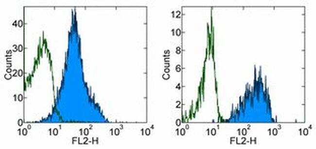
- Staining of normal human peripheral blood cells with 0.125 µg of Purified Mouse IgG1 kappa Isotype Control (Product # 14-4714-82) (open histogram) or 0.125 µg of Anti-Human CD321 (F11R) Purified (filled histogram) followed by F (ab')2 Anti-Mouse IgG PE (Product # 12-4012). Cells in the lymphocyte (left) and monocyte (right) gates were used for analysis.