 Explore
 Validate
 Learn
Learn53-3219-42
antibody from Invitrogen Antibodies
Targeting: F11R
CD321, JAM-1, JAM-A, JAM1, JAMA, JCAM, PAM-1
 Immunocytochemistry
Antibody data
- Antibody Data
- Antigen structure
- References [1]
- Comments [0]
- Validations
- Immunocytochemistry [1]
- Flow cytometry [1]
- Other assay [1]
Submit
Validation data
Reference
Comment
Report error
- Product number
- 53-3219-42 - Provider product page

- Provider
- Invitrogen Antibodies
- Product name
- CD321 (F11R) Monoclonal Antibody (CSTEM27), Alexa Fluor™ 488, eBioscience™
- Antibody type
- Monoclonal
- Antigen
- Other
- Description
- This monoclonal antibody CSTEM27 reacts with human CD321 (F11R, JAM-1, JAM-A). Applications Reported: This CSTEM27 antibody has been reported for use in immunocytochemical staining and flow cytometric analysis. This CSTEM27 antibody has been tested by immunocytochemistry of adherent cells. This can be used at less than or equal to 5 µg/mL. This CSTEM27 antibody has also been tested by flow cytometric analysis of human iPSCs. This may be used at 0.25 µg/test. A test is defined as the amount (µg) of antibody that will stain a cell sample in a final volume of 100 µL. Cell number should be determined empirically but can range from 10^5 to 10^8 cells/test. It is recommended that the antibody be carefully titrated for optimal performance in the assay of interest. Excitation: 488 nm; Emission: 519 nm; Laser: Blue Laser. Filtration: 0.2 µm post-manufacturing filtered.
- Reactivity
- Human
- Host
- Mouse
- Conjugate
- Green dye
- Isotype
- IgG
- Antibody clone number
- CSTEM27
- Vial size
- 100 Tests
- Concentration
- 5 µL/Test
- Storage
- 4° C, store in dark, DO NOT FREEZE!
Submitted references SARS-CoV-2 Spike Protein Induces Degradation of Junctional Proteins That Maintain Endothelial Barrier Integrity.
Raghavan S, Kenchappa DB, Leo MD
Frontiers in cardiovascular medicine 2021;8:687783
Frontiers in cardiovascular medicine 2021;8:687783
No comments: Submit comment
Supportive validation
- Submitted by
- Invitrogen Antibodies (provider)
- Main image

- Experimental details
- Immunocytochemistry of fixed and permeabilized human iPSCs plated on murine derived feeder cells using 5 µg/mL Mouse IgG2a K Isotype Control Alexa Fluor® 488 (Product # 53-4724-80) (left) or 5 µg/mL Anti-CD321 (F11R) Alexa Fluor® 488 (right). Nuclei are stained with DAPI. An iPSC colony (positive staining) and murine derived feeder cells (negative staining) are in both fields of view above.
- Conjugate
- Green dye

Supportive validation
- Submitted by
- Invitrogen Antibodies (provider)
- Main image
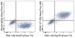
- Experimental details
- Staining of a mixture of human iPSC and the C2C12 murine cell line with Anti-Human TRA-1-60 PerCp-eFluor 710 (Product # 46-8863-82) and Mouse IgG2a K Isotype Control Alexa Fluor® 488 (Product # 53-4724-80) (left) or Anti-Human CD321 (F11R) Alexa Fluor® 488 (right).
- Conjugate
- Green dye

Supportive validation
- Submitted by
- Invitrogen Antibodies (provider)
- Main image

- Experimental details
- Figure 2 Spike increases Rab5a association with ACE2 and junctional proteins. (A) Representative Western blot of coimmunoprecipitation experiments done using Rab5a antibody and probed for different proteins. (B) Mean data indicating fold change in Rab5a association with junctional proteins and ACE2 after S1RBD treatment. n = 6 for each, * P < 0.05 vs. untreated control. (C) Representative Western blot of coimmunoprecipitation experiments done using ACE2 antibody and probed for Rab5a. (D) Mean data indicating fold change in ACE2 association with Rab5a after Spike treatment. n = 6 for each, * P < 0.05 vs. untreated control. (E) Representative Western blot of coimmunoprecipitation experiments in control and diabetic cells after Rab5a knockdown with siRNA. (F) Mean data indicating fold change in ACE2 association with Rab5a after Rab5a siRNA. n = 6 for each, * P < 0.05 vs. control, # P < 0.05 vs. NT siRNA diabetic.
- Conjugate
- Green dye
