 Explore
 Validate
 Learn
Learn710228
antibody from Invitrogen Antibodies
Targeting: F11R
CD321, JAM-1, JAM-A, JAM1, JAMA, JCAM, PAM-1
 Western blot
Antibody data
- Antibody Data
- Antigen structure
- References [0]
- Comments [0]
- Validations
- Western blot [1]
- Immunocytochemistry [1]
Submit
Validation data
Reference
Comment
Report error
- Product number
- 710228 - Provider product page

- Provider
- Invitrogen Antibodies
- Product name
- JAM-A (CD321) Recombinant Polyclonal Antibody (24HCLC)
- Antibody type
- Polyclonal
- Antigen
- Synthetic peptide
- Reactivity
- Human
- Host
- Rabbit
- Isotype
- IgG
- Antibody clone number
- 24HCLC
- Vial size
- 100 µg
- Concentration
- 0.5 mg/mL
- Storage
- Store at 4°C short term. For long term storage, store at -20°C, avoiding freeze/thaw cycles.
No comments: Submit comment
Supportive validation
- Submitted by
- Invitrogen Antibodies (provider)
- Main image

- Experimental details
- Western blot analysis of JAM1 in whole cell extracts of serum-starved MCF-7 cells treated with IGF1 (150 ng/mL, 15 min) using a JAM1 Recombinant Rabbit Polyclonal Antibody (Product # 710228) at a dilution of 2 µg/mL. Samples were detected using chemiluminescence (ECL). Results show a band at ~33 kDa.

Supportive validation
- Submitted by
- Invitrogen Antibodies (provider)
- Main image
- Experimental details
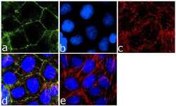
- Immunofluorescence analysis of JAM1/CD321 was done on 90% confluent log phase Caco-2 cells. The cells were fixed with 4% paraformaldehyde for 10 minutes, permeabilized with 0.1% Triton™ X-100 for 10 minutes, and blocked with 1% BSA for 1 hour at room temperature. The cells were labeled with JAM1 (24HCLC) Recombinant Rabbit Polyclonal Antibody (Product # 710228) at 2 µg/mL in 0.1% BSA and incubated for 3 hours at room temperature and then labeled with Goat anti-Rabbit IgG (H+L) Superclonal™ Secondary Antibody, Alexa Fluor® 488 conjugate (Product # A27034) at a dilution of 1:2000 for 45 minutes at room temperature (Panel a: green). Nuclei (Panel b: blue) were stained with SlowFade® Gold Antifade Mountant with DAPI (Product # S36938). F-actin (Panel c: red) was stained with Alexa Fluor® 555 Rhodamine Phalloidin (Product # R415, 1:300). Panel d is a merged image showing cell junctional localization. Panel e is a no primary antibody control. The images were captured at 60X magnification.
