Explore
 Validate
 Learn
Learn Immunocytochemistry
Antibody data
- Antibody Data
- Antigen structure
- References [1]
- Comments [0]
- Validations
- Immunocytochemistry [1]
- Immunohistochemistry [1]
- Flow cytometry [1]
Submit
Validation data
Reference
Comment
Report error
- Product number
- MAB1103 - Provider product page

- Provider
- R&D Systems
- Product name
- Human JAM-A Antibody
- Antibody type
- Monoclonal
- Description
- Protein A or G purified from hybridoma culture supernatant. Detects human JAM-A in direct ELISAs. In direct ELISAs, no cross-reactivity with recombinant human (rh) JAM-B, recombinant mouse (rm) JAM-A, or rmJAM-4 is observed.
- Reactivity
- Human
- Host
- Mouse
- Conjugate
- Unconjugated
- Antigen sequence
Q9Y624- Isotype
- IgG
- Antibody clone number
- 654806
- Vial size
- 100 ug
- Concentration
- LYOPH
- Storage
- Use a manual defrost freezer and avoid repeated freeze-thaw cycles. 12 months from date of receipt, -20 to -70 °C as supplied. 1 month, 2 to 8 °C under sterile conditions after reconstitution. 6 months, -20 to -70 °C under sterile conditions after reconstitution.
Submitted references IL-18 Is Involved in Eosinophil-Mediated Tumoricidal Activity against a Colon Carcinoma Cell Line by Upregulating LFA-1 and ICAM-1.
Gatault S, Delbeke M, Driss V, Sarazin A, Dendooven A, Kahn JE, Lefèvre G, Capron M
Journal of immunology (Baltimore, Md. : 1950) 2015 Sep 1;195(5):2483-92
Journal of immunology (Baltimore, Md. : 1950) 2015 Sep 1;195(5):2483-92
No comments: Submit comment
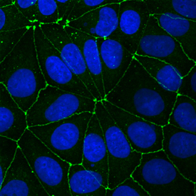
Supportive validation
- Submitted by
- R&D Systems (provider)
- Main image
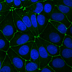
- Experimental details
- JAM-A in MCF-7 Human Cell Line. JAM-A was detected in immersion fixed MCF-7 human breast cancer cell line using Mouse Anti-Human JAM-A Monoclonal Antibody (Catalog # MAB1103) at 20 µg/mL for 3 hours at room temperature. Cells were stained using the NorthernLights™ 493-conjugated Anti-Mouse IgG Secondary Antibody (green; Catalog # NL009) and counterstained with DAPI (blue). Specific staining was localized to intercellular junctions. View our protocol for Fluorescent ICC Staining of Cells on Coverslips.
Supportive validation
- Submitted by
- R&D Systems (provider)
- Main image
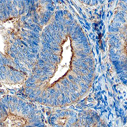
- Experimental details
- JAM-A in Human Endometrial Cancer Tissue. JAM-A was detected in immersion fixed paraffin-embedded sections of human endometrial cancer tissue using Human JAM-A Monoclonal Antibody (Catalog # MAB1103) at 15 µg/mL overnight at 4 °C. Before incubation with the primary antibody, tissue was subjected to heat-induced epitope retrieval using Antigen Retrieval Reagent-Basic (Catalog # CTS013). Tissue was stained using the Anti-Mouse HRP-DAB Cell & Tissue Staining Kit (brown; Catalog # CTS002) and counterstained with hematoxylin (blue). Specific staining was localized to the plasma membrane of glandular epithelial cells. View our protocol for Chromogenic IHC Staining of Paraffin-embedded Tissue Sections.

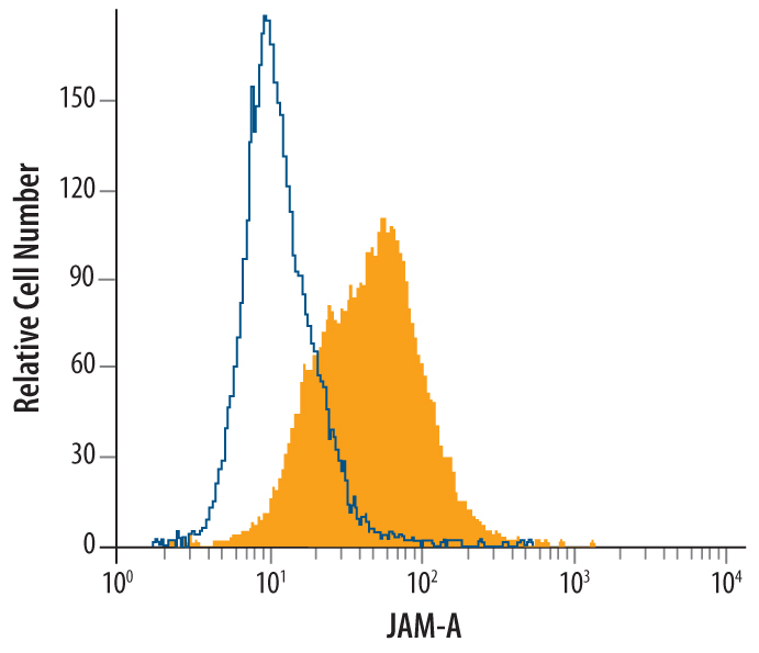
Supportive validation
- Submitted by
- R&D Systems (provider)
- Main image

- Experimental details
- Detection of JAM-A in MCF-7 Human Cell Line by Flow Cytometry. MCF-7 human breast cancer cell line was stained with Human JAM-A Monoclonal Antibody (Catalog # MAB1103, filled histogram) or isotype control antibody (Catalog # MAB002, open histogram), followed by Phycoerythrin-conjugated Anti-Mouse IgG F(ab')2 Secondary Antibody (Catalog # F0102B).