Explore
 Validate
 Learn
Learn Western blot
 Immunocytochemistry
Antibody data
- Antibody Data
- Antigen structure
- References [2]
- Comments [0]
- Validations
- Immunocytochemistry [4]
- Immunohistochemistry [4]
- Flow cytometry [2]
- Other assay [3]
Submit
Validation data
Reference
Comment
Report error
- Product number
- MA5-32843 - Provider product page

- Provider
- Invitrogen Antibodies
- Product name
- PCSK9 Monoclonal Antibody (2F1)
- Antibody type
- Monoclonal
- Antigen
- Recombinant full-length protein
- Reactivity
- Human, Mouse
- Host
- Mouse
- Isotype
- IgG
- Antibody clone number
- 2F1
- Vial size
- 100 μL
- Concentration
- 1.084 mg/mL
- Storage
- Store at 4°C short term. For long term storage, store at -20°C, avoiding freeze/thaw cycles.
Submitted references Proprotein convertase subtilisin/kexin type 9 is a psoriasis-susceptibility locus that is negatively related to IL36G.
Proprotein convertase subtilisin/kexin Type 9 is required for Ahnak-mediated metastasis of melanoma into lung epithelial cells.
Merleev A, Ji-Xu A, Toussi A, Tsoi LC, Le ST, Luxardi G, Xing X, Wasikowski R, Liakos W, Brüggen MC, Elder JT, Adamopoulos IE, Izumiya Y, Leal AR, Li Q, Kuzminykh NY, Kirane A, Marusina AI, Gudjonsson JE, Maverakis E
JCI insight 2022 Aug 22;7(16)
JCI insight 2022 Aug 22;7(16)
Proprotein convertase subtilisin/kexin Type 9 is required for Ahnak-mediated metastasis of melanoma into lung epithelial cells.
Suh JM, Son Y, Yoo JY, Goh Y, Seidah NG, Lee S, Bae YS
Neoplasia (New York, N.Y.) 2021 Sep;23(9):993-1001
Neoplasia (New York, N.Y.) 2021 Sep;23(9):993-1001
No comments: Submit comment
Supportive validation
- Submitted by
- Invitrogen Antibodies (provider)
- Main image

- Experimental details
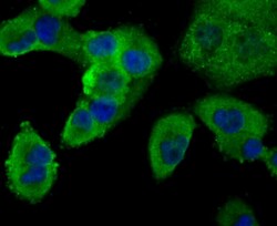
- Immunocytochemical analysis of PCSK9 in LOVO cells using a PCSK9 Monoclonal antibody (Product # MA5-32843).The nuclear counter stain is DAPI (blue). Cells were fixed in paraformaldehyde, permeabilised with 0.25% Triton X100/PBS.

- Submitted by
- Invitrogen Antibodies (provider)
- Main image
- Experimental details
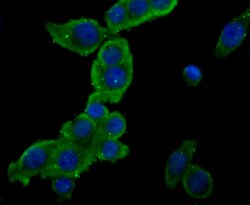
- Immunocytochemical analysis of PCSK9 in PANC-1 cells using a PCSK9 Monoclonal antibody (Product # MA5-32843).The nuclear counter stain is DAPI (blue). Cells were fixed in paraformaldehyde, permeabilised with 0.25% Triton X100/PBS.

- Submitted by
- Invitrogen Antibodies (provider)
- Main image
- Experimental details
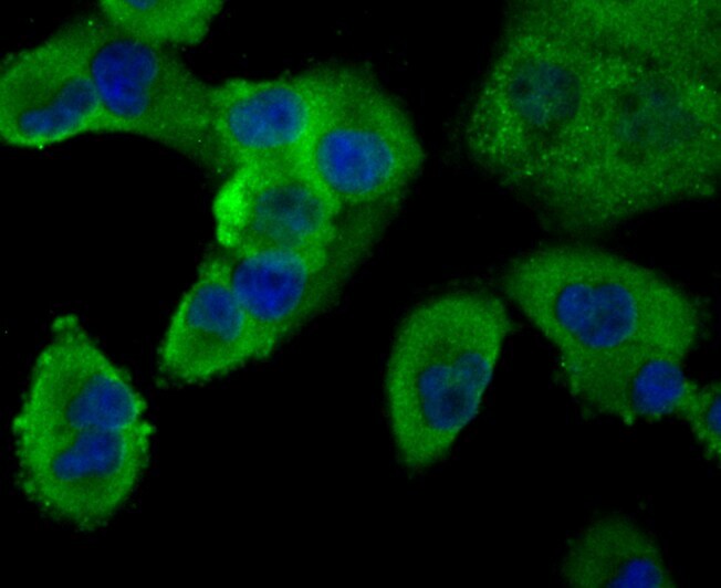
- Immunocytochemical analysis of PCSK9 in LOVO cells using a PCSK9 Monoclonal antibody (Product # MA5-32843).The nuclear counter stain is DAPI (blue). Cells were fixed in paraformaldehyde, permeabilised with 0.25% Triton X100/PBS.

- Submitted by
- Invitrogen Antibodies (provider)
- Main image

- Experimental details
- Immunocytochemical analysis of PCSK9 in PANC-1 cells using a PCSK9 Monoclonal antibody (Product # MA5-32843).The nuclear counter stain is DAPI (blue). Cells were fixed in paraformaldehyde, permeabilised with 0.25% Triton X100/PBS.
Supportive validation
- Submitted by
- Invitrogen Antibodies (provider)
- Main image
- Experimental details
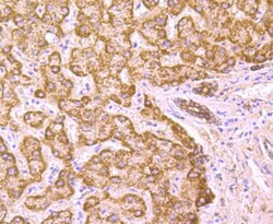
- Immunohistochemical analysis of PCSK9 of paraffin-embedded Human liver tissue using a PCSK9 Monoclonal antibody (Product #MA5-32843). Counter stained with hematoxylin.

- Submitted by
- Invitrogen Antibodies (provider)
- Main image
- Experimental details
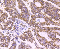
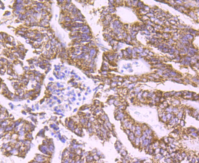
- Immunohistochemical analysis of PCSK9 of paraffin-embedded Human colon caner tissue using a PCSK9 Monoclonal antibody (Product #MA5-32843). Counter stained with hematoxylin.
- Submitted by
- Invitrogen Antibodies (provider)
- Main image
- Experimental details
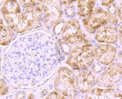
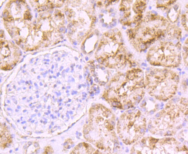
- Immunohistochemical analysis of PCSK9 of paraffin-embedded Human kidney tissue using a PCSK9 Monoclonal antibody (Product #MA5-32843). Counter stained with hematoxylin.
- Submitted by
- Invitrogen Antibodies (provider)
- Main image

- Experimental details
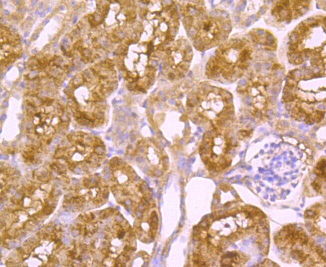
- Immunohistochemical analysis of PCSK9 of paraffin-embedded Mouse kidney tissue using a PCSK9 Monoclonal antibody (Product #MA5-32843). Counter stained with hematoxylin.
Supportive validation
- Submitted by
- Invitrogen Antibodies (provider)
- Main image

- Experimental details
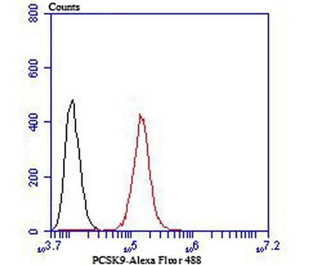
- Flow Cytometric analysis of PCSK9 in Hela cells using a PCSK9 Monoclonal Antibody (Product # MA5-32843) at a dilution of 1:100, as seen in red compared with an unlabelled control (cells without incubation with primary antibody; black). Alexa Fluor 488-conjugated Goat anti mouse IgG was used as the secondary antibody.
- Submitted by
- Invitrogen Antibodies (provider)
- Main image
- Experimental details
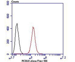
- Flow Cytometric analysis of PCSK9 in Hela cells using a PCSK9 Monoclonal Antibody (Product # MA5-32843) at a dilution of 1:100, as seen in red compared with an unlabelled control (cells without incubation with primary antibody; black). Alexa Fluor 488-conjugated Goat anti mouse IgG was used as the secondary antibody.

Supportive validation
- Submitted by
- Invitrogen Antibodies (provider)
- Main image
- Experimental details
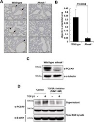
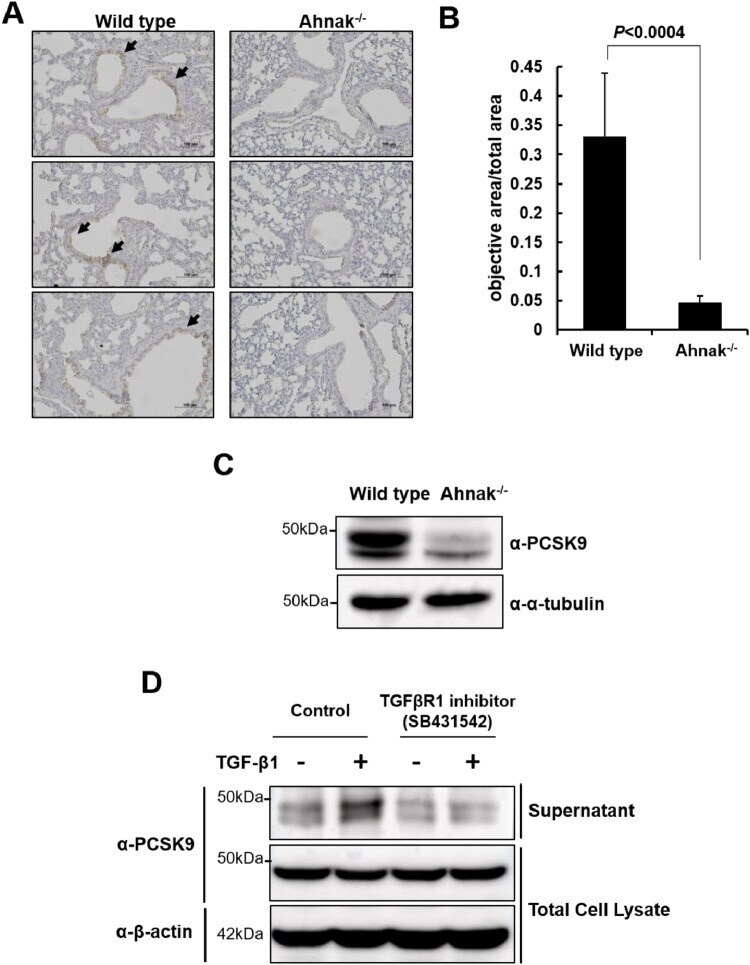
- Fig. 3 PCSK9 expression in WT and Ahnak -/- mice. (A) PCSK9 expression in the pulmonary arteries from WT and Ahnak -/- mice. Arrow means PCSK9 expression in IHC. (B) Quantitation of PCSK9 expression in (A). (C) Lysates of lung bronchial tissues from WT and Ahnak -/- mice were subjected into immunoblot analysis with antibody to Ahnak. (D) Immunoblot for PCSK9 secretion in response to TGFbeta in the absence or presence of TGFbetaR1 inhibitor. Fig 3
- Submitted by
- Invitrogen Antibodies (provider)
- Main image
- Experimental details
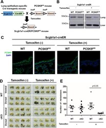
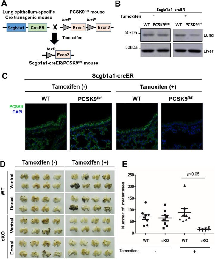
- Fig. 4 Generation and evaluation of tumor metastasis in Scgb1a1-Cre/PCSK9 fl/fl mice. (A) Strategy of generating lung epithelium-specific PCSK9 conditional KO mice. Lung epithelial cell-specific Scgb1a1 promoter-driven inducible Cre recombinase-estrogen receptor fusion protein (CreER) transgenic mouse (Scgb1a1-CreER) was crossed with Pcsk9 fl/fl mice . For PCSK9 depletion, Scgb1a1 CreER / PCSK9 fl/fl mice were intraperitoneal injected with tamoxifen (75 mg/kg) once a day for five times. (B) Lysates of lung bronchial tissues from Scgb1a1-Cre mice (control) and Scgb1a1-Cre/PCSK9 fl/fl mice were subjected into immunoblot analysis with an antibody to PCSK9. (C) Immunochemistry with antibody to PCSK9 in lung bronchial tissues from Scgb1a1-Cre and Scgb1a1-Cre/PCSK9 fl/fl mice. (D) B16F10 melanoma cells were inoculated into 8-wk-old male Scgb1a1-Cre and Scgb1a1-Cre/PCSK9 fl/fl mice via tail vein injection. The mice were sacrificed after fourteen days. The representative photograph is the dorsal side of a lung with metastatic lesions. (E) The number of metastatic colonies was counted and statistically analyzed with the Mann-Whitney U test. Fig 4
- Submitted by
- Invitrogen Antibodies (provider)
- Main image
- Experimental details
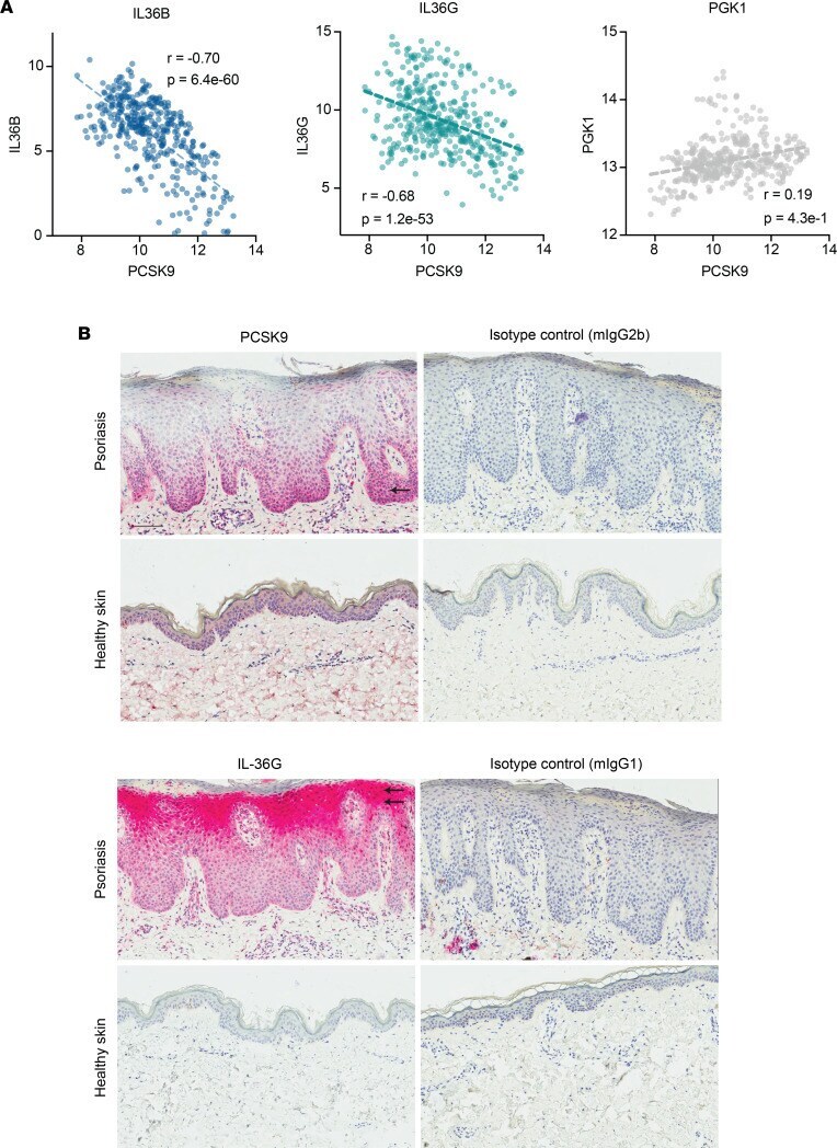
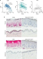
- Figure 5 PCSK9 expression negatively correlates with IL36B and IL36G expression in keratinocytes and skin. ( A ) In cultured keratinocyte cell lines, PCSK9 expression negatively correlated with IL36B and IL36G expression. PCSK9 expression did not correlate with PGK1 expression, a housekeeping gene. In these plots, each dot represents an in vitro cultured keratinocyte cell line under a different culture condition (control, IL-4, IL-13, IL-17A, IFN-alpha, IFN-gamma, TNF-alpha, IL-4 and IL-13, IL-17A and IFN-gamma, IL-17A, and TNF-alpha). Normalized log 2 transformed reads for each gene are plotted on the x axis and y axis. Pearson's correlation coefficients and P values are displayed on each plot. ( B ) Epidermal expression of PCSK9 and IL36G . Representative pictures of an IHC staining for PCSK9 and IL36G as well as the matching isotype controls in lesional skin of a patient with psoriasis (upper row) and healthy control nonlesional skin (lower row). The single arrow points to the basal layer. Double arrows point to the granular layer. Scale bar: 100 mum.