Explore
 Validate
 Learn
Learn Western blot
 ELISA
ELISAAntibody data
- Antibody Data
- Antigen structure
- References [3]
- Comments [0]
- Validations
- Western blot [2]
Submit
Validation data
Reference
Comment
Report error
- Product number
- ABIN359036 - Provider product page

- Provider
- antibodies-online
- Product name
- anti-Aurora Kinase C (AURKC) (N-Term) antibody
- Antibody type
- Polyclonal
- Antigen
- This antibody is generated from rabbits immunized with a KLH conjugated synthetic peptide selected from the N-terminal region of human Aurora-C.
- Description
- Protein G column, eluted with high and low pH buffers and neutralized immediately, followed by dialysis against PBS
- Reactivity
- Human
- Host
- Rabbit
- Epitope
- N-Term
- Vial size
- 0.4 mL
- Concentration
- 0.25 mg/mL
- Storage
- Store the antibody undiluted at 2-8°C for one month or (in aliquots) at-20°C for longer.
- Handling
- Avoid repeated freezing and thawing.
Submitted references Cell cycle-dependent expression and centrosome localization of a third human aurora/Ipl1-related protein kinase, AIK3.
Protein kinase profile of sperm and eggs: cloning and characterization of two novel testis-specific protein kinases (AIE1, AIE2) related to yeast and fly chromosome segregation regulators.
Cloning of STK13, a third human protein kinase related to Drosophila aurora and budding yeast Ipl1 that maps on chromosome 19q13.3-ter.
Kimura M, Matsuda Y, Yoshioka T, Okano Y
The Journal of biological chemistry 1999 Mar 12;274(11):7334-40
The Journal of biological chemistry 1999 Mar 12;274(11):7334-40
Protein kinase profile of sperm and eggs: cloning and characterization of two novel testis-specific protein kinases (AIE1, AIE2) related to yeast and fly chromosome segregation regulators.
Tseng TC, Chen SH, Hsu YP, Tang TK
DNA and cell biology 1998 Oct;17(10):823-33
DNA and cell biology 1998 Oct;17(10):823-33
Cloning of STK13, a third human protein kinase related to Drosophila aurora and budding yeast Ipl1 that maps on chromosome 19q13.3-ter.
Bernard M, Sanseau P, Henry C, Couturier A, Prigent C
Genomics 1998 Nov 1;53(3):406-9
Genomics 1998 Nov 1;53(3):406-9
No comments: Submit comment
Supportive validation
- Submitted by
- antibodies-online (provider)
- Main image
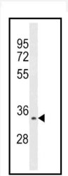
- Experimental details
- Western blot analysis of Aurora kinase C Antibody (C-term) Cat.-No AP13520PU in HepG2 cell line lysates (35ug/lane). This demonstrates the Aurora-C antibody detected the Aurora-C protein (arrow).

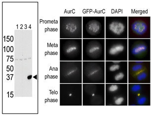
- Submitted by
- antibodies-online (provider)
- Main image
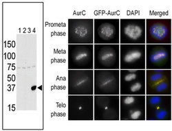
- Experimental details
- The anti-Aurora C Pab is used in Western blot to detect Aurora C in lysates of 293 cells expressing Flag tag (Lane 1), Flag-tagged Aurora A (Lane 2), Flag-tagged Aurora B (Lane 3) or Flag-tagged Aurora C (Lane 4). In the immunofluorescence experiment, staining of HeLa cells expressing GFP-Aurora C is performed at different cellular mitotic stages with the anti-Aurora C Pab as primary antibody (column A), GFP fluorescence (column B), DAPI nuclear staining (column C), and anti-Aurora C merged to DAPI staining (column D).