 Explore
 Validate
 Learn
Learn Western blot
Antibody data
- Antibody Data
- Antigen structure
- References [1]
- Comments [0]
- Validations
- Western blot [1]
- Immunocytochemistry [1]
- Flow cytometry [1]
Submit
Validation data
Reference
Comment
Report error
- Product number
- AF4649 - Provider product page

- Provider
- R&D Systems
- Product name
- Mouse Ryk Antibody
- Antibody type
- Polyclonal
- Description
- Immunogen affinity purified. Detects mouse and human Ryk in direct ELISAs and Western blots.
- Reactivity
- Mouse
- Host
- Sheep
- Conjugate
- Unconjugated
- Antigen sequence
Q01887- Isotype
- IgG
- Vial size
- 100 ug
- Concentration
- LYOPH
- Storage
- Use a manual defrost freezer and avoid repeated freeze-thaw cycles. 12 months from date of receipt, -20 to -70 °C as supplied. 1 month, 2 to 8 °C under sterile conditions after reconstitution. 6 months, -20 to -70 °C under sterile conditions after reconstitution.
Submitted references Wnt5a regulates hematopoietic stem cell proliferation and repopulation through the Ryk receptor.
Povinelli BJ, Nemeth MJ
Stem cells (Dayton, Ohio) 2014 Jan;32(1):105-15
Stem cells (Dayton, Ohio) 2014 Jan;32(1):105-15
No comments: Submit comment
Supportive validation
- Submitted by
- R&D Systems (provider)
- Main image
- Experimental details
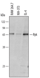
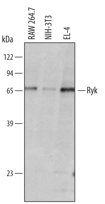
- Detection of Mouse Ryk by Western Blot. Western blot shows lysates of RAW 264.7 mouse monocyte/macrophage cell line, NIH-3T3 mouse embryonic fibroblast cell line, and EL-4 mouse lymphoblast cell line. PVDF membrane was probed with 1 µg/mL of Sheep Anti-Mouse Ryk Antigen Affinity-purified Polyclonal Antibody (Catalog # AF4649) followed by HRP-conjugated Anti-Sheep IgG Secondary Antibody (Catalog # HAF016). A specific band was detected for Ryk at approximately 70 kDa (as indicated). This experiment was conducted under reducing conditions and using Immunoblot Buffer Group 8.
Supportive validation
- Submitted by
- R&D Systems (provider)
- Main image

- Experimental details
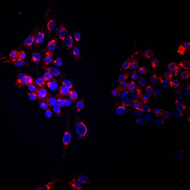
- Ryk in RAW 264.7 Mouse Cell Line. Ryk was detected in immersion fixed RAW 264.7 mouse monocyte/macrophage cell line using Sheep Anti-Mouse Ryk Antigen Affinity-purified Polyclonal Antibody (Catalog # AF4649) at 10 µg/mL for 3 hours at room temperature. Cells were stained using the NorthernLights™ 557-conjugated Anti-Sheep IgG Secondary Antibody (red; Catalog # NL010) and counterstained with DAPI (blue). Specific staining was localized to cell surfaces and cytoplasm. View our protocol for Fluorescent ICC Staining of Non-adherent Cells.
Supportive validation
- Submitted by
- R&D Systems (provider)
- Main image
- Experimental details
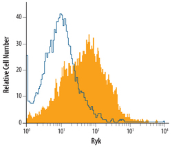
- Detection of Ryk in RAW 264.7 Mouse Cell Line by Flow Cytometry. RAW 264.7 mouse monocyte/macrophage cell line was stained with Sheep Anti-Mouse Ryk Affinity-purified Polyclonal Antibody (Catalog # AF4649, filled histogram) or isotype control antibody (Catalog # 5-001-A, open histogram), followed by Allophycocyanin-conjugated Anti-Sheep IgG Secondary Antibody (Catalog # F0127).
