Explore
 Validate
 Learn
Learn Western blot
 Immunohistochemistry
Antibody data
- Antibody Data
- Antigen structure
- References [1]
- Comments [0]
- Validations
- Immunohistochemistry [3]
- Other assay [3]
Submit
Validation data
Reference
Comment
Report error
- Product number
- PA5-105898 - Provider product page

- Provider
- Invitrogen Antibodies
- Product name
- Phospho-MEKK2 (Ser520) Polyclonal Antibody
- Antibody type
- Polyclonal
- Antigen
- Synthetic peptide
- Description
- Antibody detects endogenous levels of MAP3K2 only when phosphorylated at Ser520.
- Reactivity
- Human, Mouse, Rat
- Host
- Rabbit
- Isotype
- IgG
- Vial size
- 100 μL
- Concentration
- 1 mg/mL
- Storage
- -20°C
Submitted references MEKK2 mediates aberrant ERK activation in neurofibromatosis type I.
Bok S, Shin DY, Yallowitz AR, Eiseman M, Cung M, Xu R, Li N, Sun J, Williams AL, Scott JE, Su B, Shim JH, Greenblatt MB
Nature communications 2020 Nov 11;11(1):5704
Nature communications 2020 Nov 11;11(1):5704
No comments: Submit comment
Supportive validation
- Submitted by
- Invitrogen Antibodies (provider)
- Main image

- Experimental details
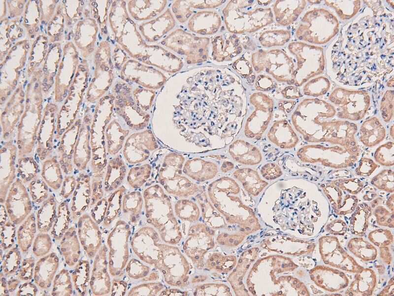
- Immunohistochemistry analysis of paraffin-embedded Phospho-MEKK2 (Ser520) in human kidney tissue sections. Antigen retrieval was performed using citrate buffer. Samples were blocked with blocking buffer (1.5 hr, 22°C), incubated with Phospho-MEKK2 (Ser520) polyclonal antibody (Product # PA5-105898) using a dilution of 1:200 (1.5 hr, 22°C), followed by HRP conjugated goat anti-rabbit.
- Submitted by
- Invitrogen Antibodies (provider)
- Main image
- Experimental details
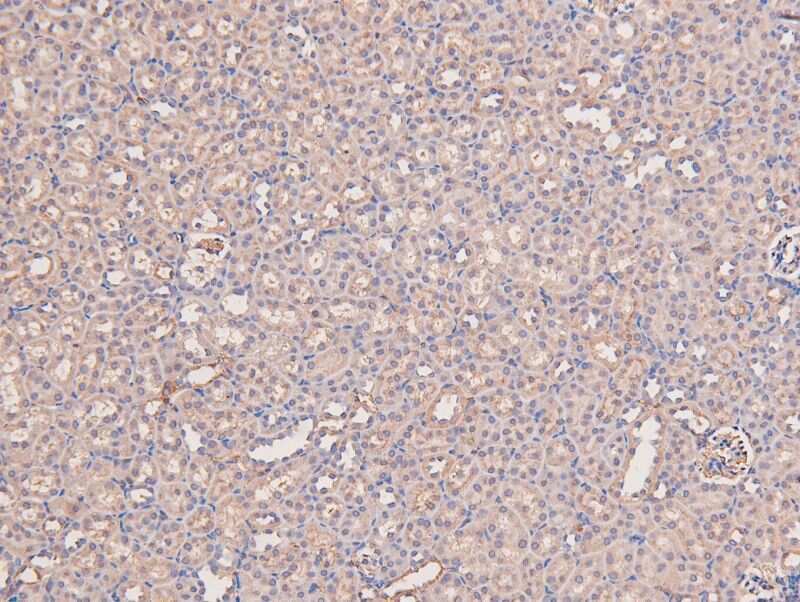
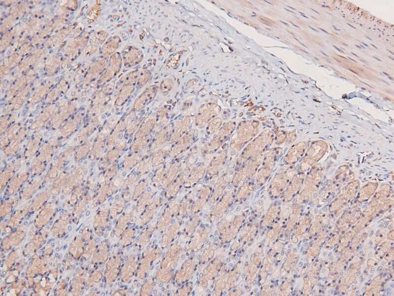
- Immunohistochemistry analysis of paraffin-embedded Phospho-MEKK2 (Ser520) in mouse kidney tissue sections. Antigen retrieval was performed using citrate buffer. Samples were blocked with blocking buffer (1.5 hr, 22°C), incubated with Phospho-MEKK2 (Ser520) polyclonal antibody (Product # PA5-105898) using a dilution of 1:200 (1.5 hr, 22°C), followed by HRP conjugated goat anti-rabbit.
- Submitted by
- Invitrogen Antibodies (provider)
- Main image
- Experimental details
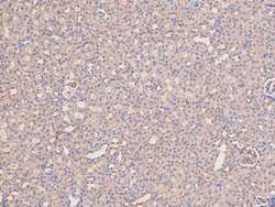
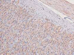
- Immunohistochemistry analysis of paraffin-embedded Phospho-MEKK2 (Ser520) in rat gastric tissue sections. Antigen retrieval was performed using citrate buffer. Samples were blocked with blocking buffer (1.5 hr, 22°C), incubated with Phospho-MEKK2 (Ser520) polyclonal antibody (Product # PA5-105898) using a dilution of 1:200 (1.5 hr, 22°C), followed by HRP conjugated goat anti-rabbit.
Supportive validation
- Submitted by
- Invitrogen Antibodies (provider)
- Main image

- Experimental details
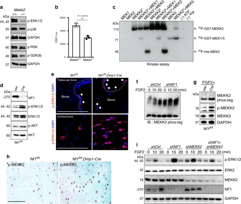
- Fig. 1 The MAP3K MEKK2 contributes to the aberrant ERK activation occurring with NF1 loss. a ERK phosphorylation levels were assessed by immunoblotting in primary COBs isolated from Mekk2 +/+ and Mekk2 -/- mice. Cell lysates were immunoblotted with the indicated antibodies. b Primary Mekk2 +/+ and Mekk2 -/- COBs were transfected with the OSE1-Luc (ATF4) reporter ( n = 3 biologically independent samples). Mean +- s.e.m., unpaired, two-tailed Student's t test: ** P < 0.01. c Purified unactivated GST-MEK1 or His-MEK2 was incubated with purified GST-MEKK2 and an in vitro kinase assay was conducted. Two types of recombinant GST-tagged MEKK2 as indicated with a dagger and double dagger were used as described in the Methods section. GST-MEK5 was used as a positive control. d Representative blot from three independent experiments. Primary Nf1 fl/fl COBs infected with either vector (Vec) or Cre lentivirus, were cultured for 7 days under differentiation conditions. Levels of NF1, phospho-ERK1/2, and ERK1/2 were analyzed by immunoblotting. e Representative images of immunostaining for p-ERK (red) in femurs from 16 weeks old Nf1 fl/fl and Nf1 fl/fl ; Dmp1-Cre mice. White arrows indicate p-ERK positive osteoblasts. Nuclei are counterstained with DAPI (blue) and scale bar indicates 100 um. Three independent fields were examined per mouse ( n = 3 mice per group). f human MSCs (hMSCs) were infected with shRNA lentiviruses expressing control (shCtrl) or NF1 (shNF1) targeting shRNAs and stimul
- Submitted by
- Invitrogen Antibodies (provider)
- Main image
- Experimental details
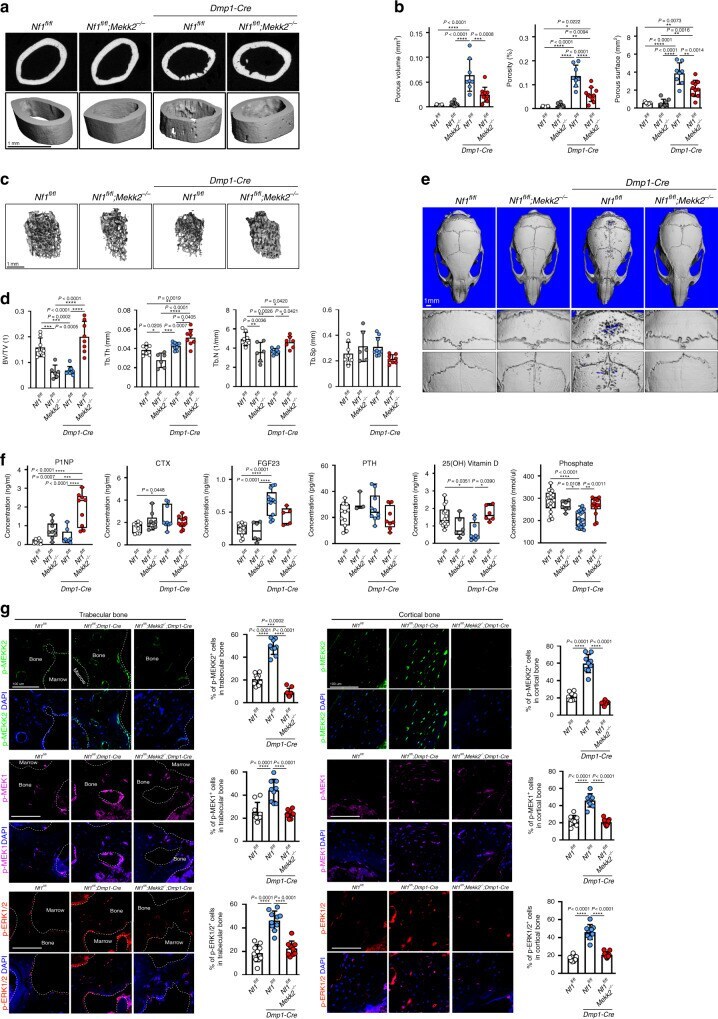
- Fig. 2 MEKK2 deficiency partially reverses NF1-associated skeletal pathology in mice. a Femurs from 16-week-old Nf1 fl/fl , Nf1 fl/fl ; Mekk2 -/- , Nf1 fl/fl ; Dmp1-Cre , and Nf1 fl/fl ; Mekk2 -/- ; Dmp1-Cre mice were analyzed by muCT. b Quantitative parameters are porous volume, porosity (porous volume/total volume), and porous surface from 16-week-old Nf1 fl/fl ( n = 5), Nf1 fl/fl ; Mekk2 -/- ( n = 8), Nf1 fl/fl ; Dmp1-Cre ( n = 8), and Nf1 fl/fl ; Mekk2 -/- ; Dmp1-Cre ( n = 10) mice. Mean +- s.d., one-way ANOVA with Tukey's multiple comparison test. c Representative muCT 3D-reconstuction images of trabecular bone in the distal femur metaphysis. d Quantitative parameters include trabecular bone volume/total volume (BV/TV), thickness (Tb.Th), trabecular number (Tb.N), and spacing (Tb.sp) in 16-week-old Nf1 fl/fl ( n = 8), Nf1 fl/fl ; Mekk2 -/- ( n = 6), Nf1 fl/fl ; Dmp1-Cre ( n = 9), and Nf1 fl/fl ; Mekk2 -/- ; Dmp1-Cre ( n = 8) mice. mean +- s.d., one-way ANOVA with Tukey's multiple comparison test. e muCT scans of mouse skulls at 16 weeks Nf1 fl/fl , Nf1 fl/fl ; Mekk2 -/- , Nf1 fl/fl ; Dmp1-Cre , and Nf1 fl/fl ; Mekk2 -/- ; Dmp1-Cre mice. f Serum levels of P1NP, CTX, FGF23, PTH, 25(OH) vitamin D, and phosphate in 16-week-old mice. Data are represented as box plots with the middle line representing the median, the box representing the 95% confidence interval of the median, and the whiskers representing the range. Each dot represents a separate mouse. g Representative images
- Submitted by
- Invitrogen Antibodies (provider)
- Main image
- Experimental details
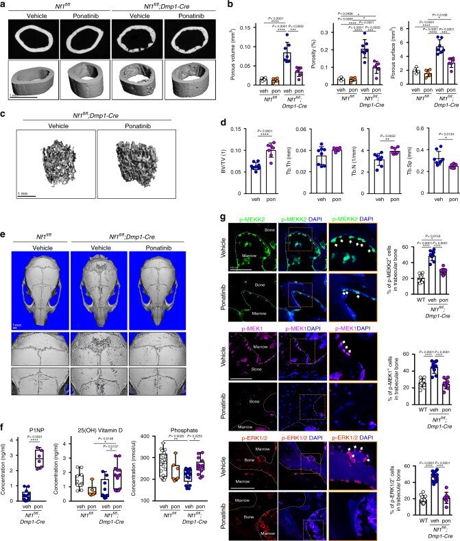
- Fig. 4 Inhibition of MEKK2 ameliorates skeletal defects in a mouse model of skeletal NF1. a Femurs from 16-week-old Nf1 fl/fl mice treated with vehicle ( n = 4) or ponatinib ( n = 5) and Nf1 fl/fl ; Dmp1-Cre mice treated with vehicle ( n = 7) or ponatinib ( n = 6) were analyzed by muCT. b Quantitative parameters are porous volume, porosity (porous volume/total volume), and porous surface from vehicle (veh) and ponatinib-treated groups (pon) of Nf1 fl/fl ;Dmp1-Cre mice. Mean +- s.d., one-way ANOVA with Tukey's multiple comparison test. c Representative muCT 3D-reconstruction images of trabecular bone in the distal femur metaphysis. d Relative quantitative analysis of trabecular BV/TV, thickness (Tb.Th), trabecular number (Tb.N), and spacing (Tb.sp) in vehicle ( n = 8) and ponatinib-treated ( n = 7) group of Nf1 fl/fl ;Dmp1-Cre . Mean +- s.d., unpaired, two-tailed Student's t test. e muCT scans of mouse skulls in 16-week-old Nf1 fl/fl and Nf1 fl/fl ;Dmp1-Cre mice treated with vehicle or ponatinib. f Serum levels of PINP, 25(OH) vitamin D, and phosphate in 16-week-old Nf1 fl/fl and Nf1 fl/fl ;Dmp1-Cre mice treated with vehicle (veh) or ponatinib (pon). Data are represented as box plots with the middle line representing the median, the box representing the 95% confidence interval of the median, and the whiskers representing the range. Each dot represents a separate mouse. g Representative immunofluorescent images and quantification for p-MEKK2 (green), p-MEK1 (magenta), and p-ERK