Explore
 Validate
 Learn
LearnMA1-23273
antibody from Invitrogen Antibodies
Targeting: RBBP4
lin-53, NURF55, RbAp48
 Western blot
 Immunocytochemistry
 Immunoprecipitation Immunohistochemistry Blocking/Neutralizing Chromatin Immunoprecipitation
Antibody data
- Antibody Data
- Antigen structure
- References [2]
- Comments [0]
- Validations
- Immunocytochemistry [3]
- Immunohistochemistry [1]
- Chromatin Immunoprecipitation [2]
Submit
Validation data
Reference
Comment
Report error
- Product number
- MA1-23273 - Provider product page

- Provider
- Invitrogen Antibodies
- Product name
- RbAp48 Monoclonal Antibody (11G10)
- Antibody type
- Monoclonal
- Antigen
- Other
- Description
- Positive control: MOLT4, HeLa, LB22. For ICC a permeabilization step is recommended; use at a dilution of 1:500-1:1000 in PBST containing 2%BSA and 10% normal serum at 4°C overnight. Store product as a concentrated solution. Centrifuge briefly prior to opening the vial.
- Reactivity
- Human, Mouse, Rat
- Host
- Mouse
- Isotype
- IgG
- Antibody clone number
- 11G10
- Vial size
- 100 μL
- Concentration
- 1 mg/mL
- Storage
- Store at 4°C short term. For long term storage, store at -20°C, avoiding freeze/thaw cycles.
Submitted references Dual retinoblastoma-binding proteins with properties related to a negative regulator of ras in yeast.
Dual retinoblastoma-binding proteins with properties related to a negative regulator of ras in yeast.
Qian YW, Lee EY
The Journal of biological chemistry 1995 Oct 27;270(43):25507-13
The Journal of biological chemistry 1995 Oct 27;270(43):25507-13
Dual retinoblastoma-binding proteins with properties related to a negative regulator of ras in yeast.
Qian YW, Lee EY
The Journal of biological chemistry 1995 Oct 27;270(43):25507-13
The Journal of biological chemistry 1995 Oct 27;270(43):25507-13
No comments: Submit comment
Supportive validation
- Submitted by
- Invitrogen Antibodies (provider)
- Main image
- Experimental details
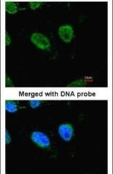
- Immunofluorescent analysis of RbAp48 in HeLa cells using a RbAp48 monoclonal antibody (Product # MA1-23273) at a 1:100 dilution.

- Submitted by
- Invitrogen Antibodies (provider)
- Main image

- Experimental details
- Immunofluorescence analysis of HeLa, using RbAp48 (Product # MA1-23273) antibody at 1:100 dilution.

- Submitted by
- Invitrogen Antibodies (provider)
- Main image
- Experimental details
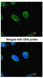
- Immunofluorescence analysis of HeLa, using RbAp48 (Product # MA1-23273) antibody at 1:100 dilution.

Supportive validation
- Submitted by
- Invitrogen Antibodies (provider)
- Main image
- Experimental details
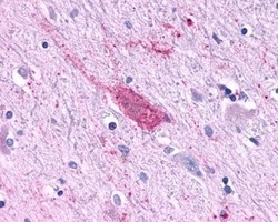
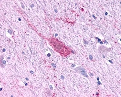
- Immunohistochemical analysis using RbAp48 Monoclonal Antibody (11G10) (Product # MA1-23273).
Supportive validation
- Submitted by
- Invitrogen Antibodies (provider)
- Main image
- Experimental details
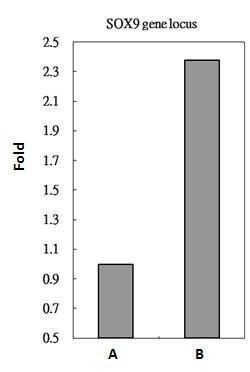
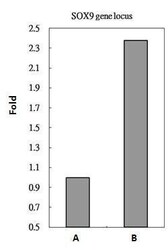
- RbAp48 antibody immunoprecipitates RbAp48 protein-DNA in ChIP experiments. ChIP Sample: HeLa whole cell lysate/extract. A: 5 µg preimmune mouse IgG. B: 5 µg of RbAp48 antibody (Product # MA1-23273). The precipitated DNA was detected by PCR with primer set targeting to SOX9 gene locus.

- Submitted by
- Invitrogen Antibodies (provider)
- Main image

- Experimental details
- RbAp48 antibody immunoprecipitates RbAp48 protein-DNA in ChIP experiments. ChIP Sample: HeLa whole cell lysate/extract. A: 5 µg preimmune mouse IgG. B: 5 µg of RbAp48 antibody (Product # MA1-23273). The precipitated DNA was detected by PCR with primer set targeting to SOX9 gene locus.