Explore
 Validate
 Learn
LearnA00244-2
antibody from Boster Biological Technology
Targeting: DDX58
DKFZp434J1111, FLJ13599, RIG-1, RIG-I, RIG1
 Western blot
 Immunocytochemistry
Antibody data
- Antibody Data
- Antigen structure
- References [0]
- Comments [0]
- Validations
- Western blot [1]
Submit
Validation data
Reference
Comment
Report error
- Product number
- A00244-2 - Provider product page

- Provider
- Boster Biological Technology
- Product name
- Anti-DDX58 Antibody Picoband™
- Antibody type
- Polyclonal
- Description
- Polyclonal antibody for RIG I/DDX58 detection. Host: Rabbit.Size: 100ug/vial. Tested applications: WB, ICC/IF, FCM. Reactive species: Human;Mouse;Rat. RIG I/DDX58 information: Molecular Weight: 106600 MW; Subcellular Localization: Cytoplasm. Cell projection, ruffle membrane. Cytoplasm, cytoskeleton. Cell junction, tight junction. Colocalized with TRIM25 at cytoplasmic perinuclear bodies. Associated with the actin cytoskeleton at membrane ruffles; Tissue Specificity: Present in vascular smooth cells (at protein level).
- Reactivity
- Human, Mouse, Rat
- Host
- Rabbit
- Vial size
- 100ug/vial
- Concentration
- Add 0.2ml of distilled water will yield a concentration of 500ug/ml.
- Storage
- At -20°C for one year. After reconstitution, at 4°C for one month. It can also be aliquoted and stored frozen at -20°C for a longer time. Avoid repeated freezing and thawing.
- Handling
- Add 0.2ml of distilled water will yield a concentration of 500ug/ml.
No comments: Submit comment
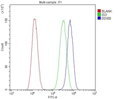
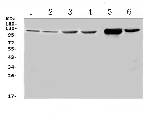
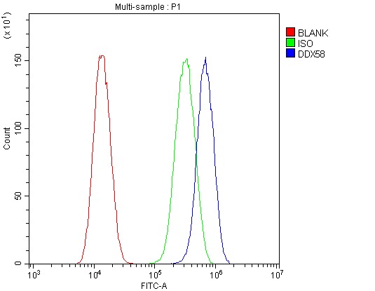
Supportive validation
- Submitted by
- Boster Biological Technology (provider)
- Main image

- Experimental details
- Western blot analysis of DDX58 using anti-DDX58 antibody (A00244-2). Electrophoresis was performed on a 5-20% SDS-PAGE gel at 70V (Stacking gel) / 90V (Resolving gel) for 2-3 hours. The sample well of each lane was loaded with 50ug of sample under reducing conditions. Lane 1: rat spleen tissue lysate,Lane 2: rat thymus tissue lysate,Lane 3: mouse spleen tissue lysate,Lane 4: mouse thymus tissue lysate,Lane 5: mouse NIH3T3 whole Cell lysate,Lane 6: human Jurkat whole Cell lysate. After Electrophoresis, proteins were transferred to a Nitrocellulose membrane at 150mA for 50-90 minutes. Blocked the membrane with 5% Non-fat Milk/ TBS for 1.5 hour at RT. The membrane was incubated with rabbit anti-DDX58 antigen affinity purified polyclonal antibody (Catalog # A00244-2) at 0.5 μg/mL overnight at 4°C, then washed with TBS-0.1%Tween 3 times with 5 minutes each and probed with a goat anti-rabbit IgG-HRP secondary antibody at a dilution of 1:10000 for 1.5 hour at RT. The signal is developed using an Enhanced Chemiluminescent detection (ECL) kit (Catalog # EK1002) with Tanon 5200 system. A specific band was detected for DDX58 at approximately 116KD. The expected band size for DDX58 is at 106KD.
- Additional image