Explore
 Validate
 Learn
LearnPA5-11726
antibody from Invitrogen Antibodies
Targeting: DDX58
DKFZp434J1111, FLJ13599, RIG-1, RIG-I, RIG1
 Western blot
Antibody data
- Antibody Data
- Antigen structure
- References [0]
- Comments [0]
- Validations
- Western blot [2]
- Immunocytochemistry [1]
Submit
Validation data
Reference
Comment
Report error
- Product number
- PA5-11726 - Provider product page

- Provider
- Invitrogen Antibodies
- Product name
- RIG-I Polyclonal Antibody
- Antibody type
- Polyclonal
- Antigen
- Synthetic peptide
- Reactivity
- Human, Mouse
- Host
- Rabbit
- Isotype
- IgG
- Vial size
- 400 µL
- Storage
- Store at 4°C short term. For long term storage, store at -20°C, avoiding freeze/thaw cycles.
No comments: Submit comment
Supportive validation
- Submitted by
- Invitrogen Antibodies (provider)
- Main image

- Experimental details
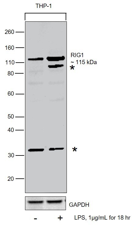
- Western blot was performed using Anti-RIG-I Polyclonal Antibody (Product # PA5-11726) and a 115 kDa band corresponding to Probable ATP-dependent RNA helicase DDX58 was observed in THP1 cells treated with 1 µg/mL LPS for 18h. Whole cell extracts (30 µg lysate) of THP-1 (Lane 1), THP-1 treated with LPS (Lane 2) were electrophoresed using NuPAGE™ 4-12% Bis-Tris Protein Gel (Product # NP0322BOX). Resolved proteins were then transferred onto a nitrocellulose membrane (Product # IB23001) by iBlot® 2 Dry Blotting System (Product # IB21001). The blot was probed with the primary antibody (1:1000) and detected by chemiluminescence with Goat anti-Rabbit IgG (H+L) Superclonal™ Recombinant Secondary Antibody, HRP (Product # A27036,1:4000) using the iBright FL 1000 (Product # A32752). Chemiluminescent detection was performed using SuperSignal™ West Dura Extended Duration Substrate (Product # 34076).
- Submitted by
- Invitrogen Antibodies (provider)
- Main image

- Experimental details
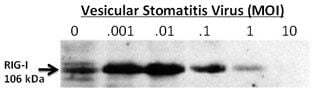
- Western blot analysis of RIG-1 in whole cell lysates from 24 hour post-infection primary murine microglia cells (2x10^6) untreated (0) or exposed to vesicular stomatitis virus at a range of viral particle/cell ratios, using a RIG-I polyclonal antibody (Product # PA5-11726).
Supportive validation
- Submitted by
- Invitrogen Antibodies (provider)
- Main image

- Experimental details
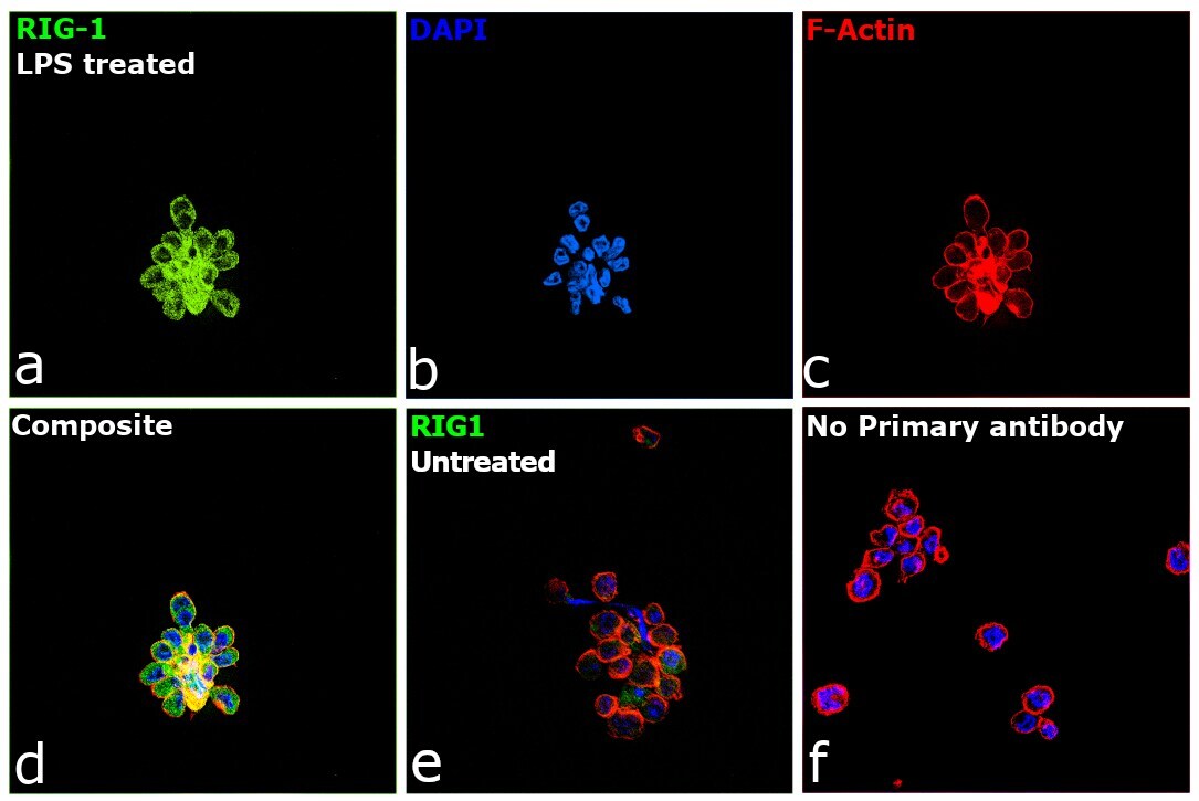
- Immunofluorescence analysis of Probable ATP-dependent RNA helicase DDX58 was performed using 70% confluent log phase THP-1 cells and THP-1 cells treated with 1 µg/mL of LPS for 18 h. The cells were fixed with 4% paraformaldehyde for 10 minutes, permeabilized with 0.1% Triton™ X-100 for 15 minutes, and blocked with 2% BSA for 45 minutes at room temperature. The cells were labeled with RIG-I Polyclonal Antibody (Product # PA5-11726) at 1:200 in 0.1% BSA, incubated at 4 degree celsius overnight and then labeled with Goat anti-Rabbit IgG (H+L) Superclonal™ Recombinant Secondary Antibody, Alexa Fluor® 488 conjugate (Product # A27034), (1:2500), for 45 minutes at room temperature (Panel a: Green). Nuclei (Panel b:Blue) were stained with ProLong™ Diamond Antifade Mountant with DAPI (Product # P36962). F-actin (Panel c: Red) was stained with Rhodamine Phalloidin (Product # R415, 1:300). Panel d represents the merged image showing Cytosolic localization. Panel f represents control cells with no primary antibody to assess background. The images were captured at 60X magnification.