 Explore
 Validate
 Learn
LearnPA5-23497
antibody from Invitrogen Antibodies
Targeting: DDX58
DKFZp434J1111, FLJ13599, RIG-1, RIG-I, RIG1
 Western blot
Antibody data
- Antibody Data
- Antigen structure
- References [0]
- Comments [0]
- Validations
- Western blot [4]
- Immunocytochemistry [1]
- Immunohistochemistry [1]
Submit
Validation data
Reference
Comment
Report error
- Product number
- PA5-23497 - Provider product page

- Provider
- Invitrogen Antibodies
- Product name
- RIG-I Polyclonal Antibody
- Antibody type
- Polyclonal
- Antigen
- Other
- Reactivity
- Human
- Host
- Rabbit
- Isotype
- IgG
- Vial size
- 100 µg
- Concentration
- 1.0 mg/mL
- Storage
- Store at 4°C short term. For long term storage, store at -20°C, avoiding freeze/thaw cycles.
No comments: Submit comment
Supportive validation
- Submitted by
- Invitrogen Antibodies (provider)
- Main image

- Experimental details
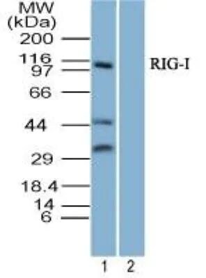
- Western blot analysis of RIG-I in 293 lysate in the 1) absence and 2) presence of immunizing peptide using a RIG-I/DDx58 polyclonal antibody (Product # PA5-23497) at 3 µg/mL. Goat anti-rabbit Ig HRP secondary antibody and an ECL substrate solution.

- Submitted by
- Invitrogen Antibodies (provider)
- Main image

- Experimental details
- Western blot analysis of RIG-I in 293 lysate in the 1) absence and 2) presence of immunizing peptide. Samples were incubated in RIG-I polyclonal antibody (Product # PA5-23497) using a dilution of 3 µg/mL followed by a goat anti-rabbit Ig HRP secondary antibody. PicoTect ECL substrate solution was used for this test.
- Submitted by
- Invitrogen Antibodies (provider)
- Main image

- Experimental details
- Knockout of RIG-I was achieved by CRISPR-Cas9 genome editing using LentiArray™ Lentiviral sgRNA (Product # A32042, Assay ID CRISPR874602_LV) and LentiArray Cas9 Lentivirus (Product # A32064). Western blot analysis of RIG-I was performed by loading 30 µg of THP-1 wild type (Lane 1), THP-1 wild type treated with 1 µg/mL LPS for 18 hours (Lane 2),THP-1 Cas9 (Lane 3), THP-1 Cas9 treated with 1 µg/mL LPS for 18 hours (Lane 4), THP-1 RIG-I KO (Lane 5) and THP-1 RIG-I KO treated with 1 µg/mL LPS for 18 hours (Lane 6) whole cell extracts. The samples were electrophoresed using NuPAGE™ Novex™ 4-12% Bis-Tris Protein Gel (Product # NP0322BOX). Resolved proteins were then transferred onto a nitrocellulose membrane (Product # IB23001) by iBlot® 2 Dry Blotting System (Product # IB21001). The blot was probed with RIG-I Polyclonal Antibody (Product # PA5-23497, 1:500 dilution) and Goat anti-Rabbit IgG (H+L) Superclonal™ Recombinant Secondary Antibody, HRP (Product # A27036, 1:5000 dilution) using the iBright™ FL 1500 (Product # A44115). Chemiluminescent detection was performed using SuperSignal™ West Dura Extended Duration Substrate (Product # 34076). Loss of signal upon CRISPR mediated knockout (KO) using the LentiArray™ CRISPR product line confirms that antibody is specific to RIG-I.

- Submitted by
- Invitrogen Antibodies (provider)
- Main image

- Experimental details
- Western blot was performed using anti-RIG-I Polyclonal Antibody (Product # PA5-23497) and a 120 kDa band corresponding to RIG-I was observed upon Lipopolysaccharide treatment in THP-1. Membrane enriched extracts (30 µg lysate) of THP-1 (Lane 1) and THP-1 treated with LPS (1 ug for 18 Hours (Lane 2)were electrophoresed using Novex® NuPAGE® 4-12 % Bis-Tris gel (Product # NP0322BOX). Resolved proteins were then transferred onto a nitrocellulose membrane (Product # IB23001) by iBlot® 2 Dry Blotting System (Product # IB21001). The blot was probed with the primary antibody (2 µg/ml) and detected by chemiluminescence with Goat anti-Rabbit IgG (H+L) Superclonal™ Recombinant Secondary Antibody, HRP conjugate (Product # A27036, 1:4000 dilution) using the iBright FL 1000 (Product # A32752). Chemiluminescent detection was performed using Novex® ECL Chemiluminescent Substrate Reagent Kit (Product # WP20005).

Supportive validation
- Submitted by
- Invitrogen Antibodies (provider)
- Main image

- Experimental details
- Immunofluorescence analysis of RIG-I was performed using 70% confluent log phase THP-1 cells and THP-1 cells treated with 1 µg of LPS for 18 hr. The cells were fixed with 4% paraformaldehyde for 10 minutes, permeabilized with 0.1% Triton™ X-100 for 15 minutes, and blocked with 2% BSA for 1 hour at room temperature. The cells were labeled with RIG-I Polyclonal Antibody (Product # PA5-23497) at 5 µg/mL in 0.1% BSA, incubated at 4 degree celsius overnight and then with Goat anti-Rabbit IgG (H+L) Highly Cross-Adsorbed Secondary Antibody, Alexa Fluor Plus 488 (Product # A32731) at a dilution of 1:2000 for 45 minutes at room temperature (Panel a: green). Nuclei (Panel b: blue) were stained with ProLong™ Diamond Antifade Mountant with DAPI (Product # P36962). F-actin (Panel c: red) was stained with Rhodamine Phalloidin (Product # R415, 1:300). Panel d represents the merged image showing membrane localization. Panel e shows untreated cells with weak membrane signal. Panel f represents control cells with no primary antibody to assess background. The images were captured at 60X magnification.

Supportive validation
- Submitted by
- Invitrogen Antibodies (provider)
- Main image

- Experimental details
- Immunohistochemical analysis of RIG-I in formalin-fixed, paraffin-embedded human uterus. Samples were incubated in RIG-I polyclonal antibody (Product # PA5-23497) using a dilution of 10 µg/mL.
