 Explore
 Validate
 Learn
Learn Western blot
 Immunoprecipitation
Antibody data
- Antibody Data
- Antigen structure
- References [4]
- Comments [0]
- Validations
- Western blot [2]
- Immunocytochemistry [1]
- Other assay [2]
Submit
Validation data
Reference
Comment
Report error
- Product number
- PA1-964 - Provider product page

- Provider
- Invitrogen Antibodies
- Product name
- PSMD2 Polyclonal Antibody
- Antibody type
- Polyclonal
- Antigen
- Recombinant full-length protein
- Description
- PA1-964 detects proteasome 19S subunit S2 from human cells. PA1-964 has been successfully used in Western blot and immunoprecipitation procedures. By Western blot, this antibody detects a 100 kDa protein representing proteasome 19S subunit S2 in HeLa cell lysate. This antibody detects, to a lesser extent an ~50-kDa protein which could correspond to subunit degradation product. PA1-964 antigen is recombinant human proteasome 19S subunit S2.
- Reactivity
- Human
- Host
- Rabbit
- Isotype
- IgG
- Vial size
- 100 µL
- Concentration
- 1 mg/mL
- Storage
- -20° C, Avoid Freeze/Thaw Cycles
Submitted references The Proteasome Ubiquitin Receptor hRpn13 and Its Interacting Deubiquitinating Enzyme Uch37 Are Required for Proper Cell Cycle Progression.
The capture proteasome assay: A method to measure proteasome activity in vitro.
Landscape of the PARKIN-dependent ubiquitylome in response to mitochondrial depolarization.
Proteasomal non-catalytic subunit PSMD2 as a potential therapeutic target in association with various clinicopathologic features in lung adenocarcinomas.
Randles L, Anchoori RK, Roden RB, Walters KJ
The Journal of biological chemistry 2016 Apr 15;291(16):8773-83
The Journal of biological chemistry 2016 Apr 15;291(16):8773-83
The capture proteasome assay: A method to measure proteasome activity in vitro.
Vigneron N, Abi Habib J, Van den Eynde BJ
Analytical biochemistry 2015 Aug 1;482:7-15
Analytical biochemistry 2015 Aug 1;482:7-15
Landscape of the PARKIN-dependent ubiquitylome in response to mitochondrial depolarization.
Sarraf SA, Raman M, Guarani-Pereira V, Sowa ME, Huttlin EL, Gygi SP, Harper JW
Nature 2013 Apr 18;496(7445):372-6
Nature 2013 Apr 18;496(7445):372-6
Proteasomal non-catalytic subunit PSMD2 as a potential therapeutic target in association with various clinicopathologic features in lung adenocarcinomas.
Matsuyama Y, Suzuki M, Arima C, Huang QM, Tomida S, Takeuchi T, Sugiyama R, Itoh Y, Yatabe Y, Goto H, Takahashi T
Molecular carcinogenesis 2011 Apr;50(4):301-9
Molecular carcinogenesis 2011 Apr;50(4):301-9
No comments: Submit comment
Supportive validation
- Submitted by
- Invitrogen Antibodies (provider)
- Main image

- Experimental details
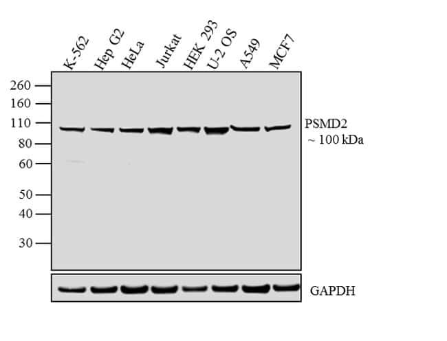
- Western blot analysis was performed on whole-cell extracts (30 µg lysate) of K-562 (Lane 1), Hep G2 (Lane 2), HeLa (Lane 3), Jurkat (Lane 4), HEK-293 (Lane 5), U-2 OS (Lane 6), A549 (Lane 7), and MCF7 (Lane 8). The blot was probed with Rabbit Anti-PSMD2 Polyclonal Antibody (Product # PA1-964, 1:500 dilution) and detected by chemiluminescence using Goat anti-Rabbit IgG (H+L) Superclonal™ Secondary Antibody, HRP conjugate (Product # A27036, 0.25 µg/mL, 1:4000 dilution). A 100 kDa band corresponding to PSMD2 was observed across the cell lines tested. Known quantity of protein samples were electrophoresed using Novex® NuPAGE® 4-12 % Bis-Tris gel (Product # NP0321BOX), XCell SureLock™ Electrophoresis System (Product # EI0002) and Novex® Sharp Pre-Stained Protein Standard (Product # LC5800). Resolved proteins were then transferred onto a nitrocellulose membrane with iBlot® 2 Dry Blotting System (Product # IB21001). The membrane was probed with the relevant primary and secondary Antibody following blocking with 5 % skimmed milk. Chemiluminescent detection was performed using Pierce™ ECL Western Blotting Substrate (Product # 32106).
- Submitted by
- Invitrogen Antibodies (provider)
- Main image
- Experimental details
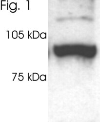
- Western blot of proteasome 19S subunit S2 on HeLa cell lysate using Product # PA1-964.

Supportive validation
- Submitted by
- Invitrogen Antibodies (provider)
- Main image
- Experimental details
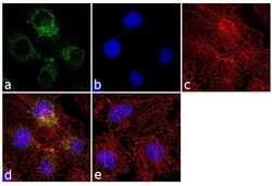
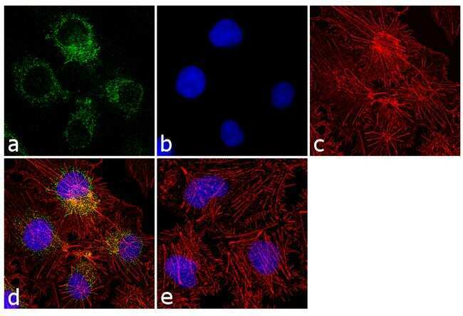
- Immunofluorescence analysis of PSMD2 was performed using 70% confluent log phase Hep G2 cells. The cells were fixed with 4% paraformaldehyde for 10 minutes, permeabilized with 0.1% Triton™ X-100 for 10 minutes, and blocked with 1% BSA for 1 hour at room temperature. The cells were labeled with PSMD2 Rabbit Polyclonal Antibody (Product # PA1-964) at 1:250 dilution in 0.1% BSA and incubated for 3 hours at room temperature and then labeled with Goat anti-Rabbit IgG (H+L) Superclonal™ Secondary Antibody, Alexa Fluor® 488 conjugate (Product # A27034) at a dilution of 1:2000 for 45 minutes at room temperature (Panel a: green). Nuclei (Panel b: blue) were stained with SlowFade® Gold Antifade Mountant with DAPI (Product # S36938). F-actin (Panel c: red) was stained with Rhodamine Phalloidin (Product # R415, 1:300). Panel d represents the merged image showing cytosolic localization. Panel e shows the control without primary antibody. The images were captured at 60X magnification.
Supportive validation
- Submitted by
- Invitrogen Antibodies (provider)
- Main image
- Experimental details
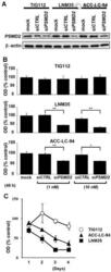
- 1 Reduction of cell growth by PSMD2 knockdown in lung cancer cell lines (LNM35 and ACC-LC-94), but not in a normal fibroblast cell line (TIG112). (A) Western blot analysis of PSMD2 at 48 h after siRNA treatment. beta-actin was used as a loading control. (B) MTT assay of cells treated with either 1 or 10 nM siRNA for 48 h. * and ** indicate the significant growth suppression with the P values of P < 0.05 and P < 0.01, respectively. (C) Results of MTT assay that measured cell growth for up to 4 days after treatment with 10 nM siRNA. OD values with si CTRL in (B) and (C) were regarded as 100%. si PSMD2 , siRNA against PSMD2 ; si CNTL , negative control siRNA.

- Submitted by
- Invitrogen Antibodies (provider)
- Main image
- Experimental details
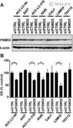
- 3 Reduction of cell growth by PSMD2 knockdown in panel of lung cancer cell lines. (A) Western blot analysis of PSMD2 in cells treated with 10 nM siRNA for 48 h. beta-actin was used as a loading control. (B) Results of MTT assay showing varying degrees of cell growth inhibition by si PSMD2 treatment. OD values with si CTRL were regarded as 100%. * and ** indicate the significant growth suppression with the P values of P < 0.05 and P < 0.01, respectively.
