 Explore
 Validate
 Learn
Learn Western blot
Antibody data
- Antibody Data
- Antigen structure
- References [2]
- Comments [0]
- Validations
- Western blot [2]
- Immunocytochemistry [1]
Submit
Validation data
Reference
Comment
Report error
- Product number
- AF3428 - Provider product page

- Provider
- R&D Systems
- Product name
- Human Lyp Antibody
- Antibody type
- Polyclonal
- Description
- Immunogen affinity purified. Detects human Lyp in Western blots.
- Reactivity
- Human
- Host
- Goat
- Conjugate
- Unconjugated
- Antigen sequence
Q9Y2R2- Isotype
- IgG
- Vial size
- 100 ug
- Concentration
- LYOPH
- Storage
- Use a manual defrost freezer and avoid repeated freeze-thaw cycles. 12 months from date of receipt, -20 to -70 °C as supplied. 1 month, 2 to 8 °C under sterile conditions after reconstitution. 6 months, -20 to -70 °C under sterile conditions after reconstitution.
Submitted references PTPN22.6, a dominant negative isoform of PTPN22 and potential biomarker of rheumatoid arthritis.
Autoimmune-associated PTPN22 R620W variation reduces phosphorylation of lymphoid phosphatase on an inhibitory tyrosine residue.
Chang HH, Tai TS, Lu B, Iannaccone C, Cernadas M, Weinblatt M, Shadick N, Miaw SC, Ho IC
PloS one 2012;7(3):e33067
PloS one 2012;7(3):e33067
Autoimmune-associated PTPN22 R620W variation reduces phosphorylation of lymphoid phosphatase on an inhibitory tyrosine residue.
Fiorillo E, Orrú V, Stanford SM, Liu Y, Salek M, Rapini N, Schenone AD, Saccucci P, Delogu LG, Angelini F, Manca Bitti ML, Schmedt C, Chan AC, Acuto O, Bottini N
The Journal of biological chemistry 2010 Aug 20;285(34):26506-18
The Journal of biological chemistry 2010 Aug 20;285(34):26506-18
No comments: Submit comment
Supportive validation
- Submitted by
- R&D Systems (provider)
- Main image

- Experimental details
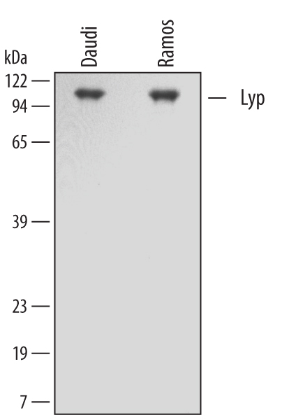
- Detection of Human Lyp by Western Blot. Western blot shows lysates of Daudi human Burkitt's lymphoma cell line and Ramos human Burkitt's lymphoma cell lines. PVDF membrane was probed with 0.3 µg/mL of Goat Anti-Human Lyp Antigen Affinity-purified Polyclonal Antibody (Catalog # AF3428) followed by HRP-conjugated Anti-Goat IgG Secondary Antibody (Catalog # HAF109). A specific band was detected for Lyp at approximately 108 kDa (as indicated). This experiment was conducted under reducing conditions and using Immunoblot Buffer Group 1.
- Submitted by
- R&D Systems (provider)
- Main image
- Experimental details
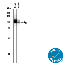
- Detection of Human Lyp by Simple WesternTM. Simple Western lane view shows lysates of Daudi human Burkitt's lymphoma cell line and Ramos human Burkitt's lymphoma cell line, loaded at 0.2 mg/mL. A specific band was detected for Lyp at approximately 116-118 kDa (as indicated) using 3 µg/mL of Goat Anti-Human Lyp Antigen Affinity-purified Polyclonal Antibody (Catalog # AF3428) followed by 1:50 dilution of HRP-conjugated Anti-Goat IgG Secondary Antibody (Catalog # HAF109). This experiment was conducted under reducing conditions and using the 12-230 kDa separation system.

Supportive validation
- Submitted by
- R&D Systems (provider)
- Main image
- Experimental details
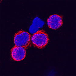
- Lyp in Human PBMCs. Lyp was detected in immersion fixed human peripheral blood mononuclear cells (PBMCs) using Goat Anti-Human Lyp Antigen Affinity-purified Polyclonal Antibody (Catalog # AF3428) at 15 µg/mL for 3 hours at room temperature. Cells were stained using the NorthernLights™ 557-conjugated Anti-Goat IgG Secondary Antibody (red; Catalog # NL001) and counterstained with DAPI (blue). Specific staining was localized to cytoplasmic. View our protocol for Fluorescent ICC Staining of Non-adherent Cells.
