Explore
 Validate
 Learn
Learn Western blot
Antibody data
- Antibody Data
- Antigen structure
- References [1]
- Comments [0]
- Validations
- Western blot [1]
- Immunohistochemistry [1]
Submit
Validation data
Reference
Comment
Report error
- Product number
- MAB3428 - Provider product page

- Provider
- R&D Systems
- Product name
- Human Lyp Antibody
- Antibody type
- Monoclonal
- Description
- Protein A or G purified from hybridoma culture supernatant. Detects human Lyp in Western blots.
- Reactivity
- Human
- Host
- Mouse
- Conjugate
- Unconjugated
- Antigen sequence
Q9Y2R2- Isotype
- IgG
- Antibody clone number
- 340113
- Vial size
- 100 ug
- Concentration
- LYOPH
- Storage
- Use a manual defrost freezer and avoid repeated freeze-thaw cycles. 12 months from date of receipt, -20 to -70 °C as supplied. 1 month, 2 to 8 °C under sterile conditions after reconstitution. 6 months, -20 to -70 °C under sterile conditions after reconstitution.
Submitted references Cutting edge: the PTPN22 allelic variant associated with autoimmunity impairs B cell signaling.
Arechiga AF, Habib T, He Y, Zhang X, Zhang ZY, Funk A, Buckner JH
Journal of immunology (Baltimore, Md. : 1950) 2009 Mar 15;182(6):3343-7
Journal of immunology (Baltimore, Md. : 1950) 2009 Mar 15;182(6):3343-7
No comments: Submit comment
Supportive validation
- Submitted by
- R&D Systems (provider)
- Main image
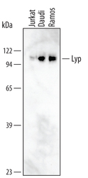
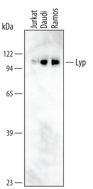
- Experimental details
- Detection of Human Lyp by Western Blot. Western blot shows lysates of Jurkat human acute T cell leukemia cell line, Daudi human Burkitt's lymphoma cell line, and Ramos human Burkitt's lymphoma cell line. PVDF membrane was probed with 1 µg/mL of Mouse Anti-Human Lyp Monoclonal Antibody (Catalog # MAB3428) followed by HRP-conjugated Anti-Mouse IgG Secondary Antibody (Catalog # HAF007). A specific band was detected for Lyp at approximately 108 kDa (as indicated). This experiment was conducted under reducing conditions and using Immunoblot Buffer Group 1.
Supportive validation
- Submitted by
- R&D Systems (provider)
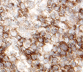
- Main image

- Experimental details
- Lyp in Human Lymph Node. Lyp was detected in immersion fixed paraffin-embedded sections of human lymph node using 25 µg/mL Mouse Anti-Human Lyp Monoclonal Antibody (Catalog # MAB3428) overnight at 4 °C. Tissue was stained with the Anti-Mouse HRP-DAB Cell & Tissue Staining Kit (brown; Catalog # CTS002) and counterstained with hematoxylin (blue). View our protocol for Chromogenic IHC Staining of Paraffin-embedded Tissue Sections.