Explore
 Validate
 Learn
Learn Western blot
 Immunocytochemistry
Antibody data
- Antibody Data
- Antigen structure
- References [11]
- Comments [0]
- Validations
- Immunocytochemistry [5]
- Other assay [1]
Submit
Validation data
Reference
Comment
Report error
- Product number
- PA1-052 - Provider product page

- Provider
- Invitrogen Antibodies
- Product name
- Leptin Polyclonal Antibody
- Antibody type
- Polyclonal
- Antigen
- Synthetic peptide
- Description
- PA1-052 detects leptin from human, mouse, and porcine samples. PA1-052 has been successfully used in Western blot and ICC/IF procedures. By Western blot, this antibody detects an ~16 kDa protein representing leptin from human serum. The PA1-052 immunogen is a synthetic peptide corresponding to residues S(91) R N V I Q I S N D L E N L R D(106) of human Leptin. This sequence is 94% conserved between human and bovine.
- Reactivity
- Human, Mouse, Porcine
- Host
- Rabbit
- Isotype
- IgG
- Vial size
- 100 μL
- Storage
- -20°C, Avoid Freeze/Thaw Cycles
Submitted references An ameloblastin C-terminus variant is present in human adipose tissue.
Expression of leptin and its long form receptor at the porcine maternal-fetal interface: contrasting healthy and arresting conceptus attachment sites during early and mid-pregnancy.
The effect of leptin on luteal angiogenic factors during the luteal phase of the estrous cycle in goats.
In vitro effects of luteinizing hormone, progesterone and oestradiol-17β on leptin gene expression and leptin secretion by porcine luteal cells obtained in early pregnancy.
Direct in vitro effect of LH and steroids on leptin gene expression and leptin secretion by porcine luteal cells during the mid-luteal phase of the estrous cycle.
IL-6 regulates exercise and training-induced adaptations in subcutaneous adipose tissue in mice.
Human skeletal muscle releases leptin in vivo.
Effects of acute fasting and age on leptin and peroxisome proliferator-activated receptor gamma production relative to fat depot in immature and mature pigs.
Expression of leptin and long-form leptin-receptor proteins in porcine hypothalamus during oestrous cycle and pregnancy.
Regulated expression of the obese gene product (leptin) in white adipose tissue and 3T3-L1 adipocytes.
Leptin levels reflect body lipid content in mice: evidence for diet-induced resistance to leptin action.
Stakkestad Ø, Heyward C, Lyngstadaas SP, Medin T, Vondrasek J, Lian AM, Pezeshki G, Reseland JE
Heliyon 2018 Dec;4(12):e01075
Heliyon 2018 Dec;4(12):e01075
Expression of leptin and its long form receptor at the porcine maternal-fetal interface: contrasting healthy and arresting conceptus attachment sites during early and mid-pregnancy.
Kerr A, Kridli RT, Khalaj K, Wessels JM, Hahnel A, Tayade C
Reproductive biology and endocrinology : RB&E 2014 Sep 23;12:91
Reproductive biology and endocrinology : RB&E 2014 Sep 23;12:91
The effect of leptin on luteal angiogenic factors during the luteal phase of the estrous cycle in goats.
Wiles JR, Katchko RA, Benavides EA, O'Gorman CW, Escudero JM, Keisler DH, Stanko RL, Garcia MR
Animal reproduction science 2014 Aug;148(3-4):121-9
Animal reproduction science 2014 Aug;148(3-4):121-9
In vitro effects of luteinizing hormone, progesterone and oestradiol-17β on leptin gene expression and leptin secretion by porcine luteal cells obtained in early pregnancy.
Siawrys G, Smolinska N
Journal of physiology and pharmacology : an official journal of the Polish Physiological Society 2013 Aug;64(4):513-20
Journal of physiology and pharmacology : an official journal of the Polish Physiological Society 2013 Aug;64(4):513-20
Direct in vitro effect of LH and steroids on leptin gene expression and leptin secretion by porcine luteal cells during the mid-luteal phase of the estrous cycle.
Siawrys G, Smolinska N
Reproductive biology 2012 Nov;12(3):317-23
Reproductive biology 2012 Nov;12(3):317-23
IL-6 regulates exercise and training-induced adaptations in subcutaneous adipose tissue in mice.
Brandt C, Jakobsen AH, Adser H, Olesen J, Iversen N, Kristensen JM, Hojman P, Wojtaszewski JF, Hidalgo J, Pilegaard H
Acta physiologica (Oxford, England) 2012 Jun;205(2):224-35
Acta physiologica (Oxford, England) 2012 Jun;205(2):224-35
Human skeletal muscle releases leptin in vivo.
Wolsk E, Mygind H, Grøndahl TS, Pedersen BK, van Hall G
Cytokine 2012 Dec;60(3):667-73
Cytokine 2012 Dec;60(3):667-73
Effects of acute fasting and age on leptin and peroxisome proliferator-activated receptor gamma production relative to fat depot in immature and mature pigs.
O'Gorman CW, Stanko RL, Keisler DH, Garcia MR
Journal of animal physiology and animal nutrition 2010 Dec;94(6):e266-76
Journal of animal physiology and animal nutrition 2010 Dec;94(6):e266-76
Expression of leptin and long-form leptin-receptor proteins in porcine hypothalamus during oestrous cycle and pregnancy.
Siawrys G, Kaminski T, Smolinska N, Przala J
Reproduction in domestic animals = Zuchthygiene 2009 Dec;44(6):920-6
Reproduction in domestic animals = Zuchthygiene 2009 Dec;44(6):920-6
Regulated expression of the obese gene product (leptin) in white adipose tissue and 3T3-L1 adipocytes.
MacDougald OA, Hwang CS, Fan H, Lane MD
Proceedings of the National Academy of Sciences of the United States of America 1995 Sep 26;92(20):9034-7
Proceedings of the National Academy of Sciences of the United States of America 1995 Sep 26;92(20):9034-7
Leptin levels reflect body lipid content in mice: evidence for diet-induced resistance to leptin action.
Frederich RC, Hamann A, Anderson S, Löllmann B, Lowell BB, Flier JS
Nature medicine 1995 Dec;1(12):1311-4
Nature medicine 1995 Dec;1(12):1311-4
No comments: Submit comment
Supportive validation
- Submitted by
- Invitrogen Antibodies (provider)
- Main image
- Experimental details
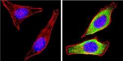
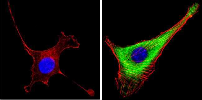
- Immunofluorescent analysis of Leptin (green) showing staining in the secretion of HeLa cells. Formalin-fixed cells were permeabilized with 0.1% Triton X-100 in TBS for 5-10 minutes and blocked with 3% BSA-PBS for 30 minutes at room temperature. Cells were probed with a Leptin polyclonal antibody (Product # PA1-052) in 3% BSA-PBS at a dilution of 1:100 and incubated overnight at 4 ºC in a humidified chamber. Cells were washed with PBST and incubated with a DyLight-conjugated secondary antibody in PBS at room temperature in the dark. F-actin (red) was stained with a fluorescent red phalloidin and nuclei (blue) were stained with Hoechst or DAPI. Images were taken at a magnification of 100x.
- Submitted by
- Invitrogen Antibodies (provider)
- Main image

- Experimental details
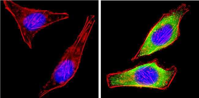
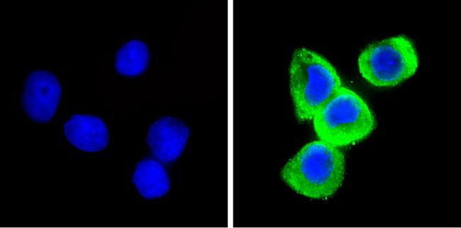
- Immunofluorescent analysis of Leptin (green) showing staining in the secretion of HepG2 cells. Formalin-fixed cells were permeabilized with 0.1% Triton X-100 in TBS for 5-10 minutes and blocked with 3% BSA-PBS for 30 minutes at room temperature. Cells were probed with a Leptin polyclonal antibody (Product # PA1-052) in 3% BSA-PBS at a dilution of 1:100 and incubated overnight at 4 ºC in a humidified chamber. Cells were washed with PBST and incubated with a DyLight-conjugated secondary antibody in PBS at room temperature in the dark. F-actin (red) was stained with a fluorescent red phalloidin and nuclei (blue) were stained with Hoechst or DAPI. Images were taken at a magnification of 100x.
- Submitted by
- Invitrogen Antibodies (provider)
- Main image

- Experimental details
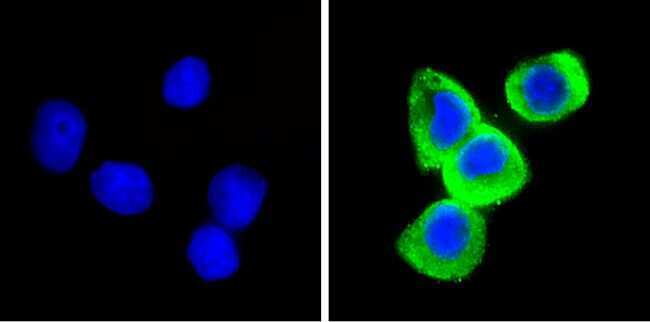
- Immunofluorescent analysis of Leptin (green) showing staining in the secretion of NIH-3T3 cells. Formalin-fixed cells were permeabilized with 0.1% Triton X-100 in TBS for 5-10 minutes and blocked with 3% BSA-PBS for 30 minutes at room temperature. Cells were probed with a Leptin polyclonal antibody (Product # PA1-052) in 3% BSA-PBS at a dilution of 1:100 and incubated overnight at 4 ºC in a humidified chamber. Cells were washed with PBST and incubated with a DyLight-conjugated secondary antibody in PBS at room temperature in the dark. F-actin (red) was stained with a fluorescent red phalloidin and nuclei (blue) were stained with Hoechst or DAPI. Images were taken at a magnification of 100x.
- Submitted by
- Invitrogen Antibodies (provider)
- Main image

- Experimental details
- Immunofluorescent analysis of Leptin (green) showing staining in the secretion of HepG2 cells. Formalin-fixed cells were permeabilized with 0.1% Triton X-100 in TBS for 5-10 minutes and blocked with 3% BSA-PBS for 30 minutes at room temperature. Cells were probed with a Leptin polyclonal antibody (Product # PA1-052) in 3% BSA-PBS at a dilution of 1:100 and incubated overnight at 4 ºC in a humidified chamber. Cells were washed with PBST and incubated with a DyLight-conjugated secondary antibody in PBS at room temperature in the dark. F-actin (red) was stained with a fluorescent red phalloidin and nuclei (blue) were stained with Hoechst or DAPI. Images were taken at a magnification of 100x.
- Submitted by
- Invitrogen Antibodies (provider)
- Main image

- Experimental details
- Immunofluorescent analysis of Leptin (green) showing staining in the secretion of NIH-3T3 cells. Formalin-fixed cells were permeabilized with 0.1% Triton X-100 in TBS for 5-10 minutes and blocked with 3% BSA-PBS for 30 minutes at room temperature. Cells were probed with a Leptin polyclonal antibody (Product # PA1-052) in 3% BSA-PBS at a dilution of 1:100 and incubated overnight at 4 ºC in a humidified chamber. Cells were washed with PBST and incubated with a DyLight-conjugated secondary antibody in PBS at room temperature in the dark. F-actin (red) was stained with a fluorescent red phalloidin and nuclei (blue) were stained with Hoechst or DAPI. Images were taken at a magnification of 100x.

Supportive validation
- Submitted by
- Invitrogen Antibodies (provider)
- Main image
- Experimental details
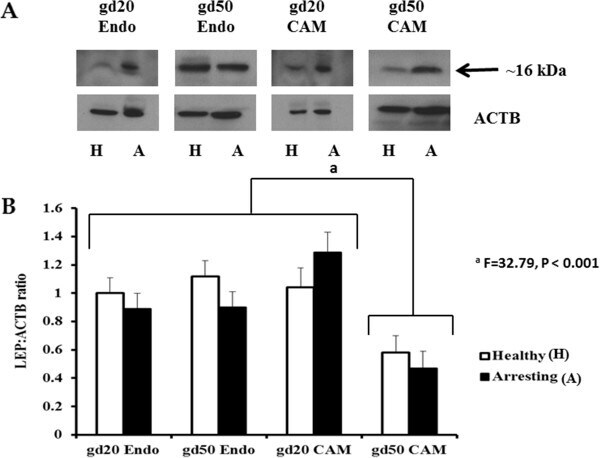
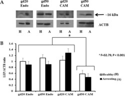
- Figure 3 Leptin (LEP) protein expression at gestational days (gd) 20 and 50 healthy and arresting conceptus attachment sites. A : Western Blotting images of LEP protein expression levels in the endometrial (Endo) and chorioallantoic membrane (CAM) tissues of healthy and arresting attachment sites at gestational day (gd) 20 and gd50. The band in the 16 kDa range was quantified relative to the expression of beta-actin (ACTB). B : Histogram represents densitometry values of relative LEP protein expression levels in the Endo and CAM tissues of healthy (white-filled bars) and arresting (black-filled bars) attachment sites at gd20 and gd50. Statistical comparisons were made based on the gd by tissue type interaction. The letter ""a"" denotes significantly lower (P < 0.001) LEP production in gd50 CAM samples than the remaining tissues at both gestational days. Histogram bars represent group means plus standard error.