Explore
 Validate
 Learn
Learn Western blot
Antibody data
- Antibody Data
- Antigen structure
- References [3]
- Comments [0]
- Validations
- Western blot [1]
- Immunohistochemistry [1]
- Blocking/Neutralizing [1]
Submit
Validation data
Reference
Comment
Report error
- Product number
- AF398 - Provider product page

- Provider
- R&D Systems
- Product name
- Human Leptin/OB Antibody
- Antibody type
- Polyclonal
- Description
- Antigen Affinity-purified. Detects human Leptin/OB in direct ELISAs and Western blots. In direct ELISAs and Western blots, less than 25% cross-reactivity with recombinant mouse Leptin/OB is observed.
- Reactivity
- Human
- Host
- Goat
- Conjugate
- Unconjugated
- Antigen sequence
Q6NT58- Isotype
- IgG
- Vial size
- 100 ug
- Concentration
- LYOPH
- Storage
- Use a manual defrost freezer and avoid repeated freeze-thaw cycles. 12 months from date of receipt, -20 to -70 °C as supplied. 1 month, 2 to 8 °C under sterile conditions after reconstitution. 6 months, -20 to -70 °C under sterile conditions after reconstitution.
Submitted references UV-induced inhibition of adipokine production in subcutaneous fat aggravates dermal matrix degradation in human skin.
Increased leptin expression in endometriosis cells is associated with endometrial stromal cell proliferation and leptin gene up-regulation.
Increased leptin expression in endometriosis cells is associated with endometrial stromal cell proliferation and leptin gene up-regulation.
Kim EJ, Kim YK, Kim MK, Kim S, Kim JY, Lee DH, Chung JH
Scientific reports 2016 May 10;6:25616
Scientific reports 2016 May 10;6:25616
Increased leptin expression in endometriosis cells is associated with endometrial stromal cell proliferation and leptin gene up-regulation.
Wu MH, Chuang PC, Chen HM, Lin CC, Tsai SJ
Molecular human reproduction 2002 May;8(5):456-64
Molecular human reproduction 2002 May;8(5):456-64
Increased leptin expression in endometriosis cells is associated with endometrial stromal cell proliferation and leptin gene up-regulation.
Wu MH, Chuang PC, Chen HM, Lin CC, Tsai SJ
Molecular human reproduction 2002 May;8(5):456-64
Molecular human reproduction 2002 May;8(5):456-64
No comments: Submit comment
Supportive validation
- Submitted by
- R&D Systems (provider)
- Main image
- Experimental details
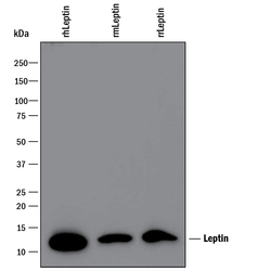
- Detection of Recombinant Human, Mouse, and Rat Leptin/OB by Western Blot. Western blot shows 100 ng of Recombinant Human Leptin/OB (Catalog # 398-LP), Recombinant Mouse Leptin/OB (Catalog # 498-OB) and Recombinant Rat Leptin/OB (Catalog # 598-LP). PVDF Membrane was probed with 0.1 µg/mL of Goat Anti-Human Leptin/OB Antigen Affinity-purified Polyclonal Antibody (Catalog # AF398) followed by HRP-conjugated Anti-Goat IgG Secondary Antibody (Catalog # HAF109). A specific band was detected for Leptin/OB at approximately 12 kDa (as indicated). This experiment was conducted under reducing conditions and using Immunoblot Buffer Group 3.

Supportive validation
- Submitted by
- R&D Systems (provider)
- Main image
- Experimental details
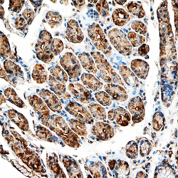
- Leptin/OB in Human Stomach. Leptin/OB was detected in immersion fixed paraffin-embedded sections of human stomach using Goat Anti-Human Leptin/OB Antigen Affinity-purified Polyclonal Antibody (Catalog # AF398) at 10 µg/mL overnight at 4 °C. Tissue was stained using the Anti-Goat HRP-DAB Cell & Tissue Staining Kit (brown; Catalog # CTS008) and counterstained with hematoxylin (blue). Specific staining was localized to cytoplasm in gastric glands. View our protocol for Chromogenic IHC Staining of Paraffin-embedded Tissue Sections.

Supportive validation
- Submitted by
- R&D Systems (provider)
- Main image
- Experimental details
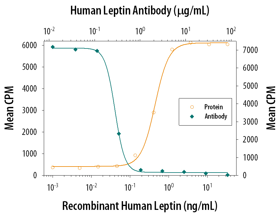
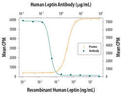
- Cell Proliferation Induced by Leptin/OB and Neutralization by Human Leptin/OB Antibody. Recombinant Human Leptin/OB (Catalog # 398-LP) stimulates proliferation in the BaF3 mouse pro-B cell line transfected with human Leptin R in a dose-dependent manner (orange line). Proliferation elicited by Recombinant Human Leptin/OB (7.5 ng/mL) is neutralized (green line) by increasing concentrations of Goat Anti-Human Leptin/OB Antigen Affinity-purified Polyclonal Antibody (Catalog # AF398). The ND50 is typically 0.5-3.0 µg/mL.