Explore
 Validate
 Learn
Learn Western blot
 Immunocytochemistry
Antibody data
- Antibody Data
- Antigen structure
- References [5]
- Comments [0]
- Validations
- Western blot [2]
Submit
Validation data
Reference
Comment
Report error
- Product number
- AF2850 - Provider product page

- Provider
- Novus Biologicals
- Product name
- Goat Polyclonal AMPK alpha 2 Antibody
- Antibody type
- Polyclonal
- Description
- Antigen Affinity-purified. Detects human, mouse, and rat AMPK alpha 2 in Western blots. In Western blots, less than 1% cross-reactivity with recombinant human AMPK alpha 1 is observed.
- Reactivity
- Human, Mouse, Rat
- Host
- Goat
- Conjugate
- Unconjugated
- Isotype
- IgG
- Vial size
- 100 ug
- Concentration
- LYOPH
- Storage
- Use a manual defrost freezer and avoid repeated freeze-thaw cycles. 12 months from date of receipt, -20 to -70 degreesC as supplied. 1 month, 2 to 8 degreesC under sterile conditions after reconstitution. 6 months, -20 to -70 degreesC under sterile conditions after reconstitution.
Submitted references Neuronal AMP-activated protein kinase hyper-activation induces synaptic loss by an autophagy-mediated process.
AMP-activated protein kinase modulates tau phosphorylation and tau pathology in vivo.
Mitochondria and AMP-activated protein kinase-dependent mechanism of efferocytosis.
Regulation of AMP-activated protein kinase signaling by AFF4 protein, member of AF4 (ALL1-fused gene from chromosome 4) family of transcription factors, in hypothalamic neurons.
The nuclear receptor PPARβ/δ programs muscle glucose metabolism in cooperation with AMPK and MEF2.
Domise M, Sauvé F, Didier S, Caillerez R, Bégard S, Carrier S, Colin M, Marinangeli C, Buée L, Vingtdeux V
Cell death & disease 2019 Mar 4;10(3):221
Cell death & disease 2019 Mar 4;10(3):221
AMP-activated protein kinase modulates tau phosphorylation and tau pathology in vivo.
Domise M, Didier S, Marinangeli C, Zhao H, Chandakkar P, Buée L, Viollet B, Davies P, Marambaud P, Vingtdeux V
Scientific reports 2016 May 27;6:26758
Scientific reports 2016 May 27;6:26758
Mitochondria and AMP-activated protein kinase-dependent mechanism of efferocytosis.
Jiang S, Park DW, Stigler WS, Creighton J, Ravi S, Darley-Usmar V, Zmijewski JW
The Journal of biological chemistry 2013 Sep 6;288(36):26013-26
The Journal of biological chemistry 2013 Sep 6;288(36):26013-26
Regulation of AMP-activated protein kinase signaling by AFF4 protein, member of AF4 (ALL1-fused gene from chromosome 4) family of transcription factors, in hypothalamic neurons.
Komori T, Doi A, Nosaka T, Furuta H, Akamizu T, Kitamura T, Senba E, Morikawa Y
The Journal of biological chemistry 2012 Jun 8;287(24):19985-96
The Journal of biological chemistry 2012 Jun 8;287(24):19985-96
The nuclear receptor PPARβ/δ programs muscle glucose metabolism in cooperation with AMPK and MEF2.
Gan Z, Burkart-Hartman EM, Han DH, Finck B, Leone TC, Smith EY, Ayala JE, Holloszy J, Kelly DP
Genes & development 2011 Dec 15;25(24):2619-30
Genes & development 2011 Dec 15;25(24):2619-30
No comments: Submit comment
Supportive validation
- Submitted by
- Novus Biologicals (provider)
- Main image
- Experimental details
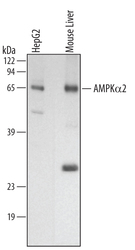
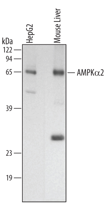
- Detection of Human and Mouse AMPK alpha 2 by Western Blot. Western blot shows lysates of HepG2 human hepatocellular carcinoma cell line and mouse liver tissue. PVDF membrane was probed with 1 µg/mL of Goat Anti-Human/Mouse/Rat AMPK alpha 2 Antigen Affinity-purified Polyclonal Antibody (Catalog # AF2850) followed by HRP-conjugated Anti-Goat IgG Secondary Antibody (Catalog # HAF109). A specific band was detected for AMPK alpha 2 at approximately 63 kDa (as indicated). This experiment was conducted under reducing conditions and using Immunoblot Buffer Group 1.
- Submitted by
- Novus Biologicals (provider)
- Main image
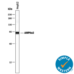
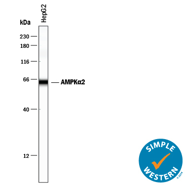
- Experimental details
- Detection of Human AMPK alpha 2 by Simple WesternTM. Simple Western lane view shows lysates of HepG2 human hepatocellular carcinoma cell line, loaded at 0.2 mg/mL. A specific band was detected for AMPK alpha 2 at approximately 63 kDa (as indicated) using 10 µg/mL of Goat Anti-Human/Mouse/Rat AMPK alpha 2 Antigen Affinity-purified Polyclonal Antibody (Catalog # AF2850) followed by 1:50 dilution of HRP-conjugated Anti-Goat IgG Secondary Antibody (Catalog # HAF109). This experiment was conducted under reducing conditions and using the 12-230 kDa separation system.