Explore
 Validate
 Learn
Learn Western blot
Antibody data
- Antibody Data
- Antigen structure
- References [0]
- Comments [0]
- Validations
- Western blot [1]
- Immunocytochemistry [1]
- Immunohistochemistry [1]
- Flow cytometry [1]
Submit
Validation data
Reference
Comment
Report error
- Product number
- MAB4260 - Provider product page

- Provider
- R&D Systems
- Product name
- Human/Mouse/Rat Jak1 Antibody
- Antibody type
- Monoclonal
- Description
- Protein A or G purified from hybridoma culture supernatant. Detects human, mouse, and rat Jak1.
- Reactivity
- Human, Mouse, Rat
- Host
- Rat
- Conjugate
- Unconjugated
- Antigen sequence
P23458- Isotype
- IgG
- Antibody clone number
- 413104
- Vial size
- 100 ug
- Concentration
- LYOPH
- Storage
- Use a manual defrost freezer and avoid repeated freeze-thaw cycles. 12 months from date of receipt, -20 to -70 °C as supplied. 1 month, 2 to 8 °C under sterile conditions after reconstitution. 6 months, -20 to -70 °C under sterile conditions after reconstitution.
No comments: Submit comment
Supportive validation
- Submitted by
- R&D Systems (provider)
- Main image
- Experimental details
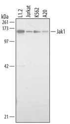
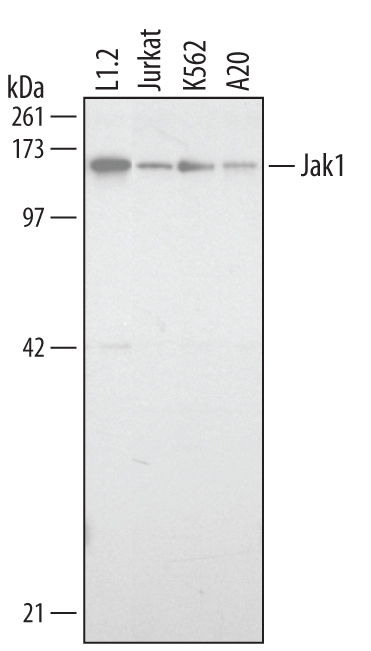
- Detection of Human and Mouse Jak1 by Western Blot. Western blot shows lysates of Jurkat human acute T cell leukemia cell line, K562 human chronic myelogenous leukemia cell line, A20 mouse B cell lymphoma cell line, and L1.2 mouse pro-B cell line. PVDF membrane was probed with 1 µg/mL of Rat Anti-Human/Mouse/Rat Jak1 Monoclonal Antibody (Catalog # MAB4260) followed by HRP-conjugated Anti-Rat IgG Secondary Antibody (Catalog # HAF005). A specific band was detected for Jak1 at approximately 130 kDa (as indicated). This experiment was conducted under reducing conditions and using Immunoblot Buffer Group 3.
Supportive validation
- Submitted by
- R&D Systems (provider)
- Main image
- Experimental details
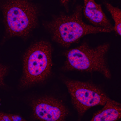
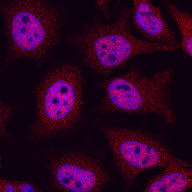
- Jak1 in HeLa Human Cell Line. Jak1 was detected in immersion fixed HeLa human cervical epithelial carcinoma cell line using Rat Anti-Human/Mouse/Rat Jak1 Monoclonal Antibody (Catalog # MAB4260) at 10 µg/mL for 3 hours at room temperature. Cells were stained using the NorthernLights™ 557-conjugated Anti-Rat IgG Secondary Antibody (red; Catalog # NL013) and counterstained with DAPI (blue). Specific staining was localized to nuclei. View our protocol for Fluorescent ICC Staining of Cells on Coverslips.
Supportive validation
- Submitted by
- R&D Systems (provider)
- Main image
- Experimental details
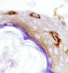
- Jak1 in Human Epidermis. Jak1 was detected in immersion fixed paraffin-embedded sections of human epidermis using 25 µg/mL Rat Anti-Human/Mouse/Rat Jak1 Monoclonal Antibody (Catalog # MAB4260) overnight at 4 °C. Tissue was stained with the Anti-Rat HRP-DAB Cell & Tissue Staining Kit (brown; Catalog # CTS017) and counterstained with hematoxylin (blue). View our protocol for Chromogenic IHC Staining of Paraffin-embedded Tissue Sections.

Supportive validation
- Submitted by
- R&D Systems (provider)
- Main image
- Experimental details
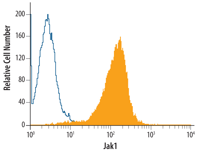
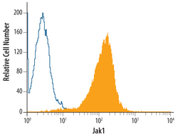
- Detection of Jak1 in Jurkat Human Cell Line by Flow Cytometry. Jurkat human acute T cell leukemia cell line was stained with Rat Anti-Human/Mouse/Rat Jak1 Monoclonal Antibody (Catalog # MAB4260, filled histogram) or isotype control antibody (Catalog # MAB0061, open histogram), followed by Phycoerythrin-conjugated Anti-Rat IgG F(ab')2 Secondary Antibody (Catalog # F0105B). To facilitate intracellular staining, cells were fixed with paraformaldehyde and permeabilized with saponin.