Explore
 Validate
 Learn
Learn Western blot
 Immunoprecipitation
Antibody data
- Antibody Data
- Antigen structure
- References [12]
- Comments [0]
- Validations
- Western blot [1]
- Immunocytochemistry [4]
- Flow cytometry [1]
- Other assay [4]
Submit
Validation data
Reference
Comment
Report error
- Product number
- PA1-2010 - Provider product page

- Provider
- Invitrogen Antibodies
- Product name
- APH1 Polyclonal Antibody
- Antibody type
- Polyclonal
- Antigen
- Synthetic peptide
- Description
- PA1-2010 detects APH-1 in human and mouse samples. PA1-2010 has been successfully used in immunofluorescent, immunoprecipitation, and Western blot procedures. By Western blot, this antibody detects a ~28 kDa protein representing APH-1 in mouse brain and kidney extracts. The PA1-2010 immunogen is a Synthetic peptide, KLH conjugated, corresponding to residues C (245) R R Q E D S R V M V Y S A L R I P P E D (265) of the C-Terminal domain of the long isoform of APH-1a. This polyclonal antibody has been referred to as, O2C2 in the reference listed above.
- Reactivity
- Human, Mouse
- Host
- Rabbit
- Isotype
- IgG
- Vial size
- 100 μg
- Concentration
- 0.69 mg/mL
- Storage
- -20°C, Avoid Freeze/Thaw Cycles
Submitted references Presenilin transmembrane domain 8 conserved AXXXAXXXG motifs are required for the activity of the γ-secretase complex.
Physical and functional interaction between the α- and γ-secretases: A new model of regulated intramembrane proteolysis.
Partial loss of presenilin impairs age-dependent neuronal survival in the cerebral cortex.
Familial frontotemporal dementia-associated presenilin-1 c.548G>T mutation causes decreased mRNA expression and reduced presenilin function in knock-in mice.
Inhibitors of γ-secretase stabilize the complex and differentially affect processing of amyloid precursor protein and other substrates.
Calsenilin regulates presenilin 1/γ-secretase-mediated N-cadherin ε-cleavage and β-catenin signaling.
Caspases-2 and -8 are involved in the presenilin1/gamma-secretase-dependent cleavage of amyloid precursor protein after the induction of apoptosis.
PI3K inhibition causes the accumulation of ubiquitinated presenilin 1 without affecting the proteasome activity.
Cell-type dependent modulation of Notch signaling by the amyloid precursor protein.
Aph-1 associates directly with full-length and C-terminal fragments of gamma-secretase substrates.
Presenilins are enriched in endoplasmic reticulum membranes associated with mitochondria.
APH-1 interacts with mature and immature forms of presenilins and nicastrin and may play a role in maturation of presenilin.nicastrin complexes.
Marinangeli C, Tasiaux B, Opsomer R, Hage S, Sodero AO, Dewachter I, Octave JN, Smith SO, Constantinescu SN, Kienlen-Campard P
The Journal of biological chemistry 2015 Mar 13;290(11):7169-84
The Journal of biological chemistry 2015 Mar 13;290(11):7169-84
Physical and functional interaction between the α- and γ-secretases: A new model of regulated intramembrane proteolysis.
Chen AC, Kim S, Shepardson N, Patel S, Hong S, Selkoe DJ
The Journal of cell biology 2015 Dec 21;211(6):1157-76
The Journal of cell biology 2015 Dec 21;211(6):1157-76
Partial loss of presenilin impairs age-dependent neuronal survival in the cerebral cortex.
Watanabe H, Iqbal M, Zheng J, Wines-Samuelson M, Shen J
The Journal of neuroscience : the official journal of the Society for Neuroscience 2014 Nov 26;34(48):15912-22
The Journal of neuroscience : the official journal of the Society for Neuroscience 2014 Nov 26;34(48):15912-22
Familial frontotemporal dementia-associated presenilin-1 c.548G>T mutation causes decreased mRNA expression and reduced presenilin function in knock-in mice.
Watanabe H, Xia D, Kanekiyo T, Kelleher RJ 3rd, Shen J
The Journal of neuroscience : the official journal of the Society for Neuroscience 2012 Apr 11;32(15):5085-96
The Journal of neuroscience : the official journal of the Society for Neuroscience 2012 Apr 11;32(15):5085-96
Inhibitors of γ-secretase stabilize the complex and differentially affect processing of amyloid precursor protein and other substrates.
Barthet G, Shioi J, Shao Z, Ren Y, Georgakopoulos A, Robakis NK
FASEB journal : official publication of the Federation of American Societies for Experimental Biology 2011 Sep;25(9):2937-46
FASEB journal : official publication of the Federation of American Societies for Experimental Biology 2011 Sep;25(9):2937-46
Calsenilin regulates presenilin 1/γ-secretase-mediated N-cadherin ε-cleavage and β-catenin signaling.
Jang C, Choi JK, Na YJ, Jang B, Wasco W, Buxbaum JD, Kim YS, Choi EK
FASEB journal : official publication of the Federation of American Societies for Experimental Biology 2011 Dec;25(12):4174-83
FASEB journal : official publication of the Federation of American Societies for Experimental Biology 2011 Dec;25(12):4174-83
Caspases-2 and -8 are involved in the presenilin1/gamma-secretase-dependent cleavage of amyloid precursor protein after the induction of apoptosis.
Chae SS, Yoo CB, Jo C, Yun SM, Jo SA, Koh YH
Journal of neuroscience research 2010 Jul;88(9):1926-33
Journal of neuroscience research 2010 Jul;88(9):1926-33
PI3K inhibition causes the accumulation of ubiquitinated presenilin 1 without affecting the proteasome activity.
Aoyagi N, Uemura K, Kuzuya A, Kihara T, Kawamata J, Shimohama S, Kinoshita A, Takahashi R
Biochemical and biophysical research communications 2010 Jan 8;391(2):1240-5
Biochemical and biophysical research communications 2010 Jan 8;391(2):1240-5
Cell-type dependent modulation of Notch signaling by the amyloid precursor protein.
Oh SY, Chen CD, Abraham CR
Journal of neurochemistry 2010 Apr;113(1):262-74
Journal of neurochemistry 2010 Apr;113(1):262-74
Aph-1 associates directly with full-length and C-terminal fragments of gamma-secretase substrates.
Chen AC, Guo LY, Ostaszewski BL, Selkoe DJ, LaVoie MJ
The Journal of biological chemistry 2010 Apr 9;285(15):11378-91
The Journal of biological chemistry 2010 Apr 9;285(15):11378-91
Presenilins are enriched in endoplasmic reticulum membranes associated with mitochondria.
Area-Gomez E, de Groof AJ, Boldogh I, Bird TD, Gibson GE, Koehler CM, Yu WH, Duff KE, Yaffe MP, Pon LA, Schon EA
The American journal of pathology 2009 Nov;175(5):1810-6
The American journal of pathology 2009 Nov;175(5):1810-6
APH-1 interacts with mature and immature forms of presenilins and nicastrin and may play a role in maturation of presenilin.nicastrin complexes.
Gu Y, Chen F, Sanjo N, Kawarai T, Hasegawa H, Duthie M, Li W, Ruan X, Luthra A, Mount HT, Tandon A, Fraser PE, St George-Hyslop P
The Journal of biological chemistry 2003 Feb 28;278(9):7374-80
The Journal of biological chemistry 2003 Feb 28;278(9):7374-80
No comments: Submit comment
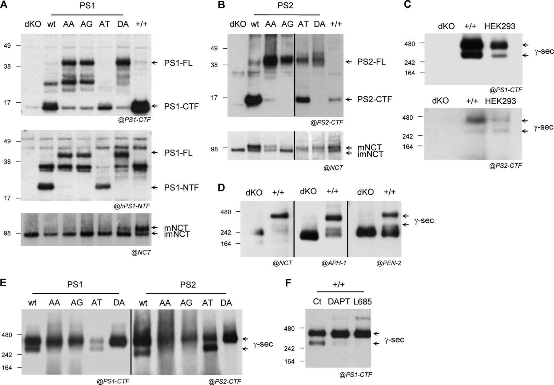
Supportive validation
- Submitted by
- Invitrogen Antibodies (provider)
- Main image

- Experimental details
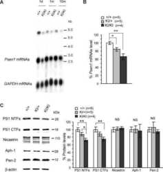
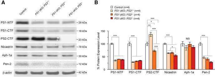
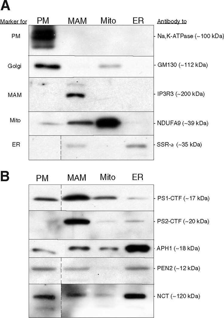
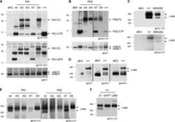
- Western blot of APH-1 in mouse brain and kidney extracts using Product # PA1-2010.
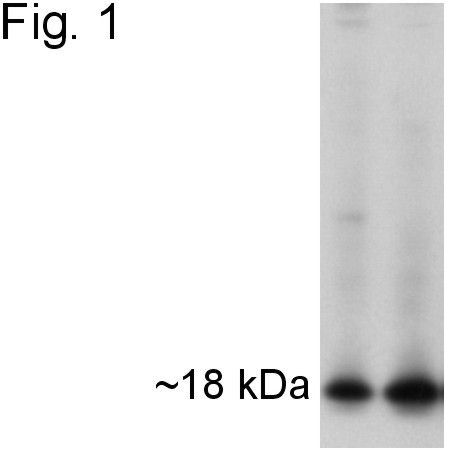
Supportive validation
- Submitted by
- Invitrogen Antibodies (provider)
- Main image
- Experimental details
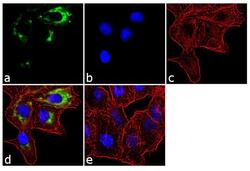
- Immunofluorescent analysis of APH-1 was performed using 70% confluent log phase A549 cells. The cells were fixed with 4% paraformaldehyde for 10 minutes, permeabilized with 0.1% Triton™ X-100 for 10 minutes, and blocked with 1% BSA for 1 hour at room temperature. The cells were labeled with APH-1 Rabbit Polyclonal Antibody (Product # PA1-2010) at 2 µg/mL in 0.1% BSA and incubated for 3 hours at room temperature and then labeled with Goat anti-Rabbit IgG (H+L) Superclonal™ Secondary Antibody, Alexa Fluor® 488 conjugate (Product # A27034) a dilution of 1:2000 for 45 minutes at room temperature (Panel a: green). Nuclei (Panel b: blue) were stained with SlowFade® Gold Antifade Mountant with DAPI (Product # S36938). F-actin (Panel c: red) was stained with Alexa Fluor® 555 Rhodamine Phalloidin (Product # R415, 1:300). Panel d represents the merged image showing cytoplasmic localization. Panel e shows the no primary antibody control. The images were captured at 60X magnification.
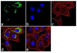
- Submitted by
- Invitrogen Antibodies (provider)
- Main image

- Experimental details
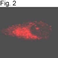
- Immunofluorescence staining of APH-1 in human KNS-42 glioma cells using Product # PA1-2010.

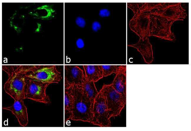
- Submitted by
- Invitrogen Antibodies (provider)
- Main image

- Experimental details
- Immunofluorescence staining of APH-1 in human KNS-42 glioma cells using Product # PA1-2010.
- Submitted by
- Invitrogen Antibodies (provider)
- Main image
- Experimental details
- Immunofluorescent analysis of APH-1 was performed using 70% confluent log phase A549 cells. The cells were fixed with 4% paraformaldehyde for 10 minutes, permeabilized with 0.1% Triton™ X-100 for 10 minutes, and blocked with 1% BSA for 1 hour at room temperature. The cells were labeled with APH-1 Rabbit Polyclonal Antibody (Product # PA1-2010) at 2 µg/mL in 0.1% BSA and incubated for 3 hours at room temperature and then labeled with Goat anti-Rabbit IgG (Heavy Chain) Superclonal™ Secondary Antibody, Alexa Fluor® 488 conjugate (Product # A27034) a dilution of 1:2000 for 45 minutes at room temperature (Panel a: green). Nuclei (Panel b: blue) were stained with SlowFade® Gold Antifade Mountant with DAPI (Product # S36938). F-actin (Panel c: red) was stained with Alexa Fluor® 555 Rhodamine Phalloidin (Product # R415, 1:300). Panel d represents the merged image showing cytoplasmic localization. Panel e shows the no primary antibody control. The images were captured at 60X magnification.

Supportive validation
- Submitted by
- Invitrogen Antibodies (provider)
- Main image
- Experimental details
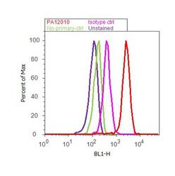
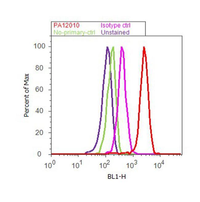
- Flow cytometry analysis of APH-1 was done on A549 cells. Cells were fixed with 70% ethanol for 10 minutes, permeabilized with 0.25% Triton™ X-100 for 20 minutes, and blocked with 5% BSA for 30 minutes at room temperature. Cells were labeled with APH-1 Rabbit Polyclonal Antibody (PA1-2010, red histogram) or with rabbit isotype control (pink histogram) at 3-5 ug/million cells in 2.5% BSA. After incubation at room temperature for 2 hours, the cells were labeled with Alexa Fluor® 488 Goat Anti-Rabbit Secondary Antibody (A11008) at a dilution of 1:400 for 30 minutes at room temperature. The representative 10, 000 cells were acquired and analyzed for each sample using an Attune® Acoustic Focusing Cytometer. The purple histogram represents unstained control cells and the green histogram represents no-primary-antibody control.
Supportive validation
- Submitted by
- Invitrogen Antibodies (provider)
- Main image
- Experimental details
- NULL

- Submitted by
- Invitrogen Antibodies (provider)
- Main image

- Experimental details
- NULL
- Submitted by
- Invitrogen Antibodies (provider)
- Main image

- Experimental details
- NULL
- Submitted by
- Invitrogen Antibodies (provider)
- Main image
- Experimental details
- NULL