 Explore
 Validate
 Learn
Learn Western blot
 Immunocytochemistry
Antibody data
- Antibody Data
- Antigen structure
- References [1]
- Comments [0]
- Validations
- Immunocytochemistry [1]
- Blocking/Neutralizing [1]
Submit
Validation data
Reference
Comment
Report error
- Product number
- AF1605 - Provider product page

- Provider
- R&D Systems
- Product name
- Equine IL-10 Antibody
- Antibody type
- Polyclonal
- Description
- Antigen Affinity-purified. Detects equine IL-10 in ELISAs and Western blots. In sandwich immunoassays, less than 1% cross-reactivity with recombinant canine IL-10 and recombinant porcine IL-10 is observed and less than 0.4% cross-reactivity with recombinant human IL-10, recombinant mouse IL-10, recombinant rat IL-10, and recombinant feline IL-10 is observed.
- Host
- Goat
- Conjugate
- Unconjugated
- Antigen sequence
Q28374- Isotype
- IgG
- Vial size
- 100 ug
- Concentration
- LYOPH
- Storage
- Use a manual defrost freezer and avoid repeated freeze-thaw cycles. 12 months from date of receipt, -20 to -70 °C as supplied. 1 month, 2 to 8 °C under sterile conditions after reconstitution. 6 months, -20 to -70 °C under sterile conditions after reconstitution.
Submitted references Attenuation of AD-like neuropathology by harnessing peripheral immune cells: local elevation of IL-10 and MMP-9.
Koronyo-Hamaoui M, Ko MK, Koronyo Y, Azoulay D, Seksenyan A, Kunis G, Pham M, Bakhsheshian J, Rogeri P, Black KL, Farkas DL, Schwartz M
Journal of neurochemistry 2009 Dec;111(6):1409-24
Journal of neurochemistry 2009 Dec;111(6):1409-24
No comments: Submit comment
Supportive validation
- Submitted by
- R&D Systems (provider)
- Main image
- Experimental details
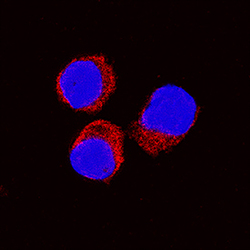
- IL-10 in Equine PBMCs. IL-10 was detected in immersion fixed equine peripheral blood mononuclear cells (PBMCs) treated with calcium ionomycin and PMA using Goat Anti-Equine IL-10 Antigen Affinity-purified Polyclonal Antibody (Catalog # AF1605) at 15 µg/mL for 3 hours at room temperature. Cells were stained using the NorthernLights™ 557-conjugated Anti-Goat IgG Secondary Antibody (red; Catalog # NL001) and counterstained with DAPI (blue). Specific staining was localized to cytoplasm. View our protocol for Fluorescent ICC Staining of Non-adherent Cells.

Supportive validation
- Submitted by
- R&D Systems (provider)
- Main image

- Experimental details
- Cell Proliferation Induced by IL-10 and Neutralization by Equine IL-10 Antibody. Recombinant Equine IL-10 (Catalog # 1605-IL) stimulates proliferation in the MC/9-2 mouse mast cell line in a dose-dependent manner (orange line). Proliferation elicited by Recombinant Equine IL-10 (20 ng/mL) is neutralized (green line) by increasing concentrations of Goat Anti-Equine IL-10 Antigen Affinity-purified Polyclonal Antibody (Catalog # AF1605). The ND50 is typically 0.2-0.6 µg/mL.
