 Explore
 Validate
 Learn
Learn Immunocytochemistry
Antibody data
- Antibody Data
- Antigen structure
- References [1]
- Comments [0]
- Validations
- Immunocytochemistry [1]
Submit
Validation data
Reference
Comment
Report error
- Product number
- MAB6932 - Provider product page

- Provider
- R&D Systems
- Product name
- Porcine IL-10 Antibody
- Antibody type
- Monoclonal
- Description
- Protein A or G purified from hybridoma culture supernatant. Detects porcine IL-10 in direct ELISAs. In direct ELISAs, 100% cross-reactivity with recombinant canine IL-10, recombinant feline IL-10, recombinant human IL-10, and recombinant viral IL-10 is observed and no cross-reactivity with recombinant cotton rat IL-10, recombinant equine IL-10, recombinant mouse IL-10, recombinant rat IL-10, or recombinant human IL-22 is observed.
- Reactivity
- Porcine
- Host
- Mouse
- Conjugate
- Unconjugated
- Antigen sequence
Q29055- Isotype
- IgG
- Antibody clone number
- 262715
- Vial size
- 100 ug
- Concentration
- LYOPH
- Storage
- Use a manual defrost freezer and avoid repeated freeze-thaw cycles. 12 months from date of receipt, -20 to -70 °C as supplied. 1 month, 2 to 8 °C under sterile conditions after reconstitution. 6 months, -20 to -70 °C under sterile conditions after reconstitution.
Submitted references Anti-inflammatory activity of nanocrystalline silver-derived solutions in porcine contact dermatitis.
Nadworny PL, Wang J, Tredget EE, Burrell RE
Journal of inflammation (London, England) 2010 Feb 19;7:13
Journal of inflammation (London, England) 2010 Feb 19;7:13
No comments: Submit comment
Supportive validation
- Submitted by
- R&D Systems (provider)
- Main image
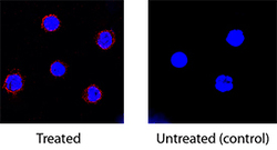
- Experimental details
- IL-10 in Porcine PBMCs. IL-10 was detected in immersion fixed porcine peripheral blood mononuclear cells treated with calcium ionomycin and PMA using Mouse Anti-Porcine IL-10 Monoclonal Antibody (Catalog # MAB6932) at 15 µg/mL for 3 hours at room temperature. Cells were stained using the NorthernLights™ 557-conjugated Anti-Mouse IgG Secondary Antibody (red; Catalog # NL007) and counterstained with DAPI (blue). Specific staining was localized to cytoplasm. View our protocol for Fluorescent ICC Staining of Non-adherent Cells.
