Explore
 Validate
 Learn
Learn Western blot
Antibody data
- Antibody Data
- Antigen structure
- References [1]
- Comments [0]
- Validations
- Western blot [1]
- Immunohistochemistry [1]
- Flow cytometry [1]
Submit
Validation data
Reference
Comment
Report error
- Product number
- AP52181PU-N - Provider product page

- Provider
- OriGene
- Product name
- IL10 (Center) rabbit polyclonal antibody, Aff - Purified
- Antibody type
- Polyclonal
- Description
- IL10 (Center) rabbit polyclonal antibody, Aff - Purified
- Host
- Rabbit
- Conjugate
- Unconjugated
- Epitope
- IL10
- Antibody clone number
- NULL
- Vial size
- 400 µl
- Concentration
- 0.5 mg/ml (Lot specific)
Submitted references Neuroinflammatory signals in Alzheimer disease and APP/PS1 transgenic mice: correlations with plaques, tangles, and oligomeric species.
López-González I, Schlüter A, Aso E, Garcia-Esparcia P, Ansoleaga B, LLorens F, Carmona M, Moreno J, Fuso A, Portero-Otin M, Pamplona R, Pujol A, Ferrer I
Journal of neuropathology and experimental neurology 2015 Apr;74(4):319-44
Journal of neuropathology and experimental neurology 2015 Apr;74(4):319-44
No comments: Submit comment
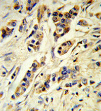
Supportive validation
- Submitted by
- OriGene (provider)
- Main image

- Experimental details
- Western blot analysis of IL10 (arrow) in MDA-MB435 cell line lysates (35ug/lane) using Interleukin-10 / IL10 antibody . .
- Validation comment
- WB
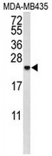
Supportive validation
- Submitted by
- OriGene (provider)
- Main image

- Experimental details
- Human breast carcinoma (formalin-fixed, paraffin-embedded) reacted with IL10 antibody .?, which was peroxidase-conjugated to the secondary antibody, followed by DAB staining. This data demonstrates the use of this antibody for IHC; clinical relevance has not been evaluated.
- Validation comment
- IHC
Supportive validation
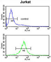
- Submitted by
- OriGene (provider)
- Main image

- Experimental details
- Flow cytometry analysis of Jurkat cells (bottom histogram) compared to a negative control cell (top histogram) using Interleukin-10/IL10 antibody . . FITC-conjugated goat-anti-rabbit secondary antibodies were used for the analysis.
- Validation comment
- FC