Explore
 Validate
 Learn
Learn Western blot
 ELISA
ELISAAntibody data
- Antibody Data
- Antigen structure
- References [2]
- Comments [0]
- Validations
- Western blot [3]
- Immunohistochemistry [2]
Submit
Validation data
Reference
Comment
Report error
- Product number
- NB100-866 - Provider product page

- Provider
- Novus Biologicals
- Proper citation
- Novus Cat#NB100-866, RRID:AB_2113148
- Product name
- Goat Polyclonal GRB2 Antibody
- Antibody type
- Polyclonal
- Description
- Immunogen affinity purified. This antibody is expected to recognize both reported isoforms (NP_002077.1and NP_987102.1).
- Reactivity
- Human, Mouse, Rat, Porcine
- Host
- Goat
- Isotype
- IgG
- Vial size
- 0.1 mg
- Concentration
- 0.5 mg/ml
- Storage
- Store at -20C. Avoid freeze-thaw cycles.
Submitted references The SH2 and SH3 domain-containing protein GRB2 links receptor tyrosine kinases to ras signaling.
The SH2 and SH3 domain-containing protein GRB2 links receptor tyrosine kinases to ras signaling.
Lowenstein EJ, Daly RJ, Batzer AG, Li W, Margolis B, Lammers R, Ullrich A, Skolnik EY, Bar-Sagi D, Schlessinger J
Cell 1992 Aug 7;70(3):431-42
Cell 1992 Aug 7;70(3):431-42
The SH2 and SH3 domain-containing protein GRB2 links receptor tyrosine kinases to ras signaling.
Lowenstein EJ, Daly RJ, Batzer AG, Li W, Margolis B, Lammers R, Ullrich A, Skolnik EY, Bar-Sagi D, Schlessinger J
Cell 1992 Aug 7;70(3):431-42
Cell 1992 Aug 7;70(3):431-42
No comments: Submit comment
Supportive validation
- Submitted by
- Novus Biologicals (provider)
- Main image
- Experimental details
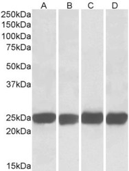
- Western Blot: GRB2 Antibody [NB100-866] - (0.05ug/ml) staining of Mouse (A+C) and Rat (B+D) Brain (A+B) and Spleen (C+D) lysates (35ug total protein in RIPA buffer). Primary incubated for 1 hour. Detected by western blot using chemiluminescence.
- Submitted by
- Novus Biologicals (provider)
- Main image

- Experimental details
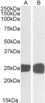
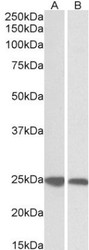
- Western Blot: GRB2 Antibody [NB100-866] - (0.05ug/ml) staining of Human Thymus (A) and Molt4 (B) lysates (35ug total protein in RIPA buffer). Primary incubated for 1 hour. Detected by western blot using chemiluminescence.
- Submitted by
- Novus Biologicals (provider)
- Main image
- Experimental details
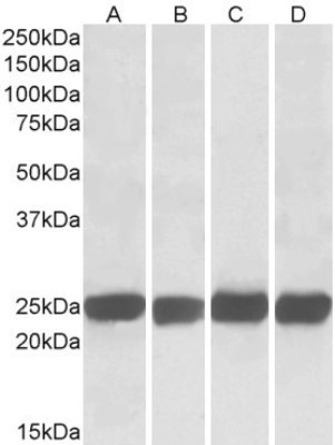
- Western Blot: GRB2 Antibody [NB100-866] - (0.03ug/ml) staining of Human (A) and Pig (B) Spleen lysates (35ug protein in RIPA buffer). Primary incubation was 1 hour. Detected by chemiluminescence.

Supportive validation
- Submitted by
- Novus Biologicals (provider)
- Main image
- Experimental details
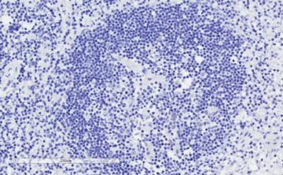
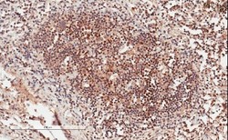
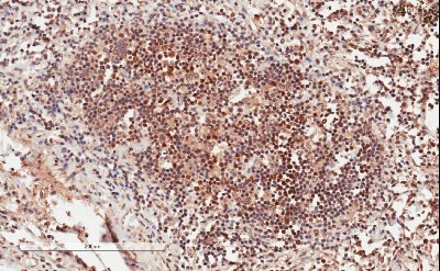
- Immunohistochemistry-Paraffin: GRB2 Antibody [NB100-866] - (1ug/ml) staining of paraffin embedded Human Lymph Node. Microwaved antigen retrieval with citrate buffer pH 6, HRP-staining.
- Submitted by
- Novus Biologicals (provider)
- Main image
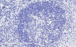
- Experimental details
- Immunohistochemistry-Paraffin: GRB2 Antibody [NB100-866] - Negative Control showing staining of paraffin embedded Human Lymph Node, with no primary antibody.