Explore
 Validate
 Learn
Learn Western blot
 Immunocytochemistry
Antibody data
- Antibody Data
- Antigen structure
- References [1]
- Comments [0]
- Validations
- Immunocytochemistry [3]
- Immunohistochemistry [6]
Submit
Validation data
Reference
Comment
Report error
- Product number
- PA5-27151 - Provider product page

- Provider
- Invitrogen Antibodies
- Product name
- GRB2 Polyclonal Antibody
- Antibody type
- Polyclonal
- Antigen
- Synthetic peptide
- Description
- Recommended positive controls: 293T, A431, HeLa, HepG2, NIH-3T3, BCL-1. Predicted reactivity: Mouse (100%), Rat (100%), Pig (100%), Chimpanzee (100%), Bovine (100%). Store product as a concentrated solution. Centrifuge briefly prior to opening the vial.
- Reactivity
- Human, Mouse, Rat
- Host
- Rabbit
- Isotype
- IgG
- Vial size
- 100 μL
- Concentration
- 1 mg/mL
- Storage
- Store at 4°C short term. For long term storage, store at -20°C, avoiding freeze/thaw cycles.
Submitted references Defactinib attenuates osteoarthritis by inhibiting positive feedback loop between H-type vessels and MSCs in subchondral bone.
Hu Y, Wu H, Xu T, Wang Y, Qin H, Yao Z, Chen P, Xie Y, Ji Z, Yang K, Chai Y, Zhang X, Yu B, Cui Z
Journal of orthopaedic translation 2020 Sep;24:12-22
Journal of orthopaedic translation 2020 Sep;24:12-22
No comments: Submit comment
Supportive validation
- Submitted by
- Invitrogen Antibodies (provider)
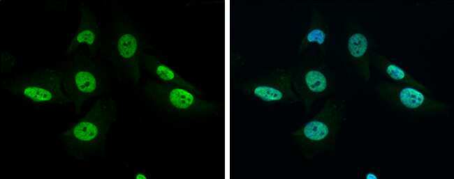
- Main image

- Experimental details
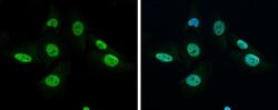
- Immunocytochemistry-Immunofluorescence analysis of GRB2 was performed in SK-N-SH cells fixed in 4% paraformaldehyde at RT for 15 min. Green: GRB2 Polyclonal Antibody (Product # PA5-27151) diluted at 1:500. Blue: Hoechst 33342 staining.
- Submitted by
- Invitrogen Antibodies (provider)
- Main image
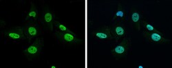
- Experimental details
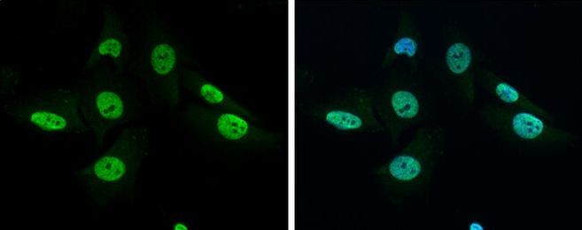
- Immunocytochemistry-Immunofluorescence analysis of GRB2 was performed in SK-N-SH cells fixed in 4% paraformaldehyde at RT for 15 min. Green: GRB2 Polyclonal Antibody (Product # PA5-27151) diluted at 1:500. Blue: Hoechst 33342 staining.
- Submitted by
- Invitrogen Antibodies (provider)
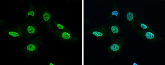
- Main image
- Experimental details
- Immunocytochemistry-Immunofluorescence analysis of GRB2 was performed in SK-N-SH cells fixed in 4% paraformaldehyde at RT for 15 min. Green: GRB2 Polyclonal Antibody (Product # PA5-27151) diluted at 1:500. Blue: Hoechst 33342 staining.
Supportive validation
- Submitted by
- Invitrogen Antibodies (provider)
- Main image
- Experimental details
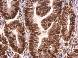
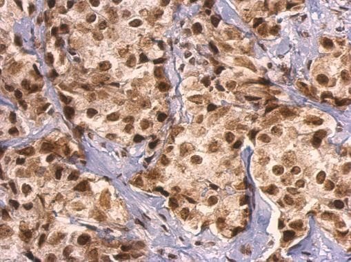
- GRB2 Polyclonal Antibody detects GRB2 protein at nucleus on human colon carcinoma by immunohistochemical analysis. Sample: Paraffin-embedded human colon carcinoma. GRB2 Polyclonal Antibody (Product # PA5-27151) dilution: 1:500. Antigen Retrieval: EDTA based buffer, pH 8.0, 15 min.

- Submitted by
- Invitrogen Antibodies (provider)
- Main image
- Experimental details
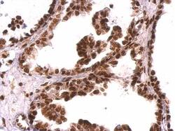
- GRB2 Polyclonal Antibody detects GRB2 protein at nucleus on human ovarian carcinoma by immunohistochemical analysis. Sample: Paraffin-embedded human ovarian carcinoma. GRB2 Polyclonal Antibody (Product # PA5-27151) dilution: 1:500. Antigen Retrieval: EDTA based buffer, pH 8.0, 15 min.

- Submitted by
- Invitrogen Antibodies (provider)
- Main image
- Experimental details
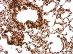
- GRB2 Polyclonal Antibody detects GRB2 protein at nucleus on mouse lung by immunohistochemical analysis. Sample: Paraffin-embedded mouse lung. GRB2 Polyclonal Antibody (Product # PA5-27151) dilution: 1:500. Antigen Retrieval: EDTA based buffer, pH 8.0, 15 min.

- Submitted by
- Invitrogen Antibodies (provider)
- Main image
- Experimental details
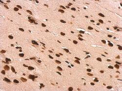
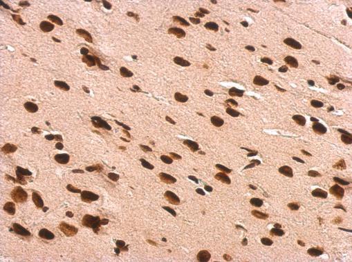
- GRB2 Polyclonal Antibody detects GRB2 protein at nucleus on rat fore brain by immunohistochemical analysis. Sample: Paraffin-embedded rat fore brain. GRB2 Polyclonal Antibody (Product # PA5-27151) dilution: 1:500. Antigen Retrieval: EDTA based buffer, pH 8.0, 15 min.
- Submitted by
- Invitrogen Antibodies (provider)
- Main image
- Experimental details
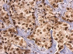
- GRB2 Polyclonal Antibody detects GRB2 protein at nucleus on human breast carcinoma by immunohistochemical analysis. Sample: Paraffin-embedded human breast carcinoma. GRB2 Polyclonal Antibody (Product # PA5-27151) dilution: 1:500. Antigen Retrieval: EDTA based buffer, pH 8.0, 15 min.
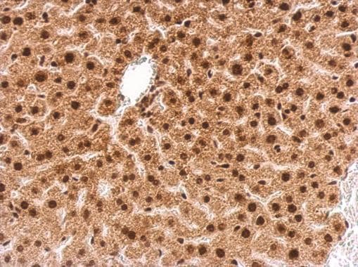
- Submitted by
- Invitrogen Antibodies (provider)
- Main image

- Experimental details
- GRB2 Polyclonal Antibody detects GRB2 protein at nucleus on mouse liver by immunohistochemical analysis. Sample: Paraffin-embedded mouse liver. GRB2 Polyclonal Antibody (Product # PA5-27151) dilution: 1:500. Antigen Retrieval: EDTA based buffer, pH 8.0, 15 min.