 Explore
 Validate
 Learn
Learn Western blot
Antibody data
- Antibody Data
- Antigen structure
- References [5]
- Comments [0]
- Validations
- Western blot [2]
- Immunohistochemistry [3]
Submit
Validation data
Reference
Comment
Report error
- Product number
- AF2685 - Provider product page

- Provider
- Novus Biologicals
- Product name
- Rabbit Polyclonal Src Antibody
- Antibody type
- Polyclonal
- Description
- Immunogen affinity purified. Detects human Src family members when phosphorylated at sites corresponding to Y419 of human Src in Western blots.
- Reactivity
- Human
- Host
- Rabbit
- Conjugate
- Unconjugated
- Isotype
- IgG
- Vial size
- 100 ug
- Concentration
- LYOPH
- Storage
- Use a manual defrost freezer and avoid repeated freeze-thaw cycles. 12 months from date of receipt, -20 to -70 degreesC as supplied. 1 month, 2 to 8 degreesC under sterile conditions after reconstitution. 6 months, -20 to -70 degreesC under sterile conditions after reconstitution.
Submitted references Clonorchis sinensis excretory-secretory products promote the migration and invasion of cholangiocarcinoma cells by activating the integrin β4-FAK/Src signaling pathway.
Role and regulation of Yap in KrasG12D-induced lung cancer.
Intravitreal AAV2.COMP-Ang1 Prevents Neurovascular Degeneration in a Murine Model of Diabetic Retinopathy.
Kaposi's sarcoma-associated herpesvirus induces rapid release of angiopoietin-2 from endothelial cells.
Targeting SRC in mucinous ovarian carcinoma.
Pak JH, Bashir Q, Kim IK, Hong SJ, Maeng S, Bahk YY, Kim TS
Molecular and biochemical parasitology 2017 Jun;214:1-9
Molecular and biochemical parasitology 2017 Jun;214:1-9
Role and regulation of Yap in KrasG12D-induced lung cancer.
Mao Y, Sun S, Irvine KD
Oncotarget 2017 Dec 19;8(67):110877-110889
Oncotarget 2017 Dec 19;8(67):110877-110889
Intravitreal AAV2.COMP-Ang1 Prevents Neurovascular Degeneration in a Murine Model of Diabetic Retinopathy.
Cahoon JM, Rai RR, Carroll LS, Uehara H, Zhang X, O'Neil CL, Medina RJ, Das SK, Muddana SK, Olson PR, Nielson S, Walker K, Flood MM, Messenger WB, Archer BJ, Barabas P, Krizaj D, Gibson CC, Li DY, Koh GY, Gao G, Stitt AW, Ambati BK
Diabetes 2015 Dec;64(12):4247-59
Diabetes 2015 Dec;64(12):4247-59
Kaposi's sarcoma-associated herpesvirus induces rapid release of angiopoietin-2 from endothelial cells.
Ye FC, Zhou FC, Nithianantham S, Chandran B, Yu XL, Weinberg A, Gao SJ
Journal of virology 2013 Jun;87(11):6326-35
Journal of virology 2013 Jun;87(11):6326-35
Targeting SRC in mucinous ovarian carcinoma.
Matsuo K, Nishimura M, Bottsford-Miller JN, Huang J, Komurov K, Armaiz-Pena GN, Shahzad MM, Stone RL, Roh JW, Sanguino AM, Lu C, Im DD, Rosenshien NB, Sakakibara A, Nagano T, Yamasaki M, Enomoto T, Kimura T, Ram PT, Schmeler KM, Gallick GE, Wong KK, Frumovitz M, Sood AK
Clinical cancer research : an official journal of the American Association for Cancer Research 2011 Aug 15;17(16):5367-78
Clinical cancer research : an official journal of the American Association for Cancer Research 2011 Aug 15;17(16):5367-78
No comments: Submit comment
Supportive validation
- Submitted by
- Novus Biologicals (provider)
- Main image

- Experimental details
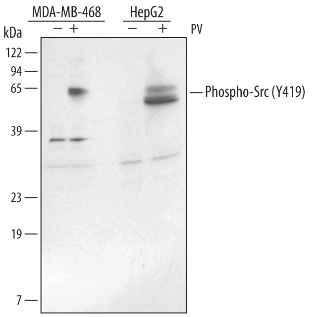
- Detection of Human Phospho-Src (Y419) by Western Blot. Western blot shows lysates of MDA-MB-468 human breast cancer cell line and HepG2 human hepatocellular carcinoma cell line untreated (-) or treated (+) with 50 μM pervanadate (PV) for 10 minutes. PVDF membrane was probed with 1 µg/mL of Rabbit Anti-Human Phospho-Src (Y419) Antigen Affinity-purified Polyclonal Antibody (Catalog # AF2685), followed by HRP-conjugated Anti-Rabbit IgG Secondary Antibody (Catalog # HAF008). A specific band was detected for Phospho-Src (Y419) at approximately 56 - 61 kDa (as indicated). This experiment was conducted under reducing conditions and using Immunoblot Buffer Group 1.
- Submitted by
- Novus Biologicals (provider)
- Main image
- Experimental details
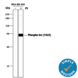
- Detection of Human Phospho-Src (Y419) by Simple WesternTM. Simple Western lane view shows lysates of MBA-MB-468 human breast cancer cell line untreated (-) or treated (+) with 50 µM Pervanadate (PV) for 10 minutes, loaded at 0.2 mg/mL. A specific band was detected for Phospho-Src (Y419) at approximately 64 kDa (as indicated) using 10 µg/mL of Rabbit Anti-Human Phospho-Src (Y419) Antigen Affinity-purified Polyclonal Antibody (Catalog # AF2685). This experiment was conducted under reducing conditions and using the 12-230 kDa separation system.

Supportive validation
- Submitted by
- Novus Biologicals (provider)
- Main image

- Experimental details
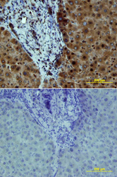
- Src in Human Liver Cancer Tissue. Src phosphorylated at Y419 was detected in immersion fixed paraffin-embedded sections of human liver cancer tissue using Rabbit Anti-Human Phospho-Src (Y419) Antigen Affinity-purified Polyclonal Antibody (Catalog # AF2685) at 10 µg/mL overnight at 4 °C. Tissue was stained using the Anti-Rabbit HRP-DAB Cell & Tissue Staining Kit (brown; Catalog # CTS005) and counterstained with hematoxylin (blue). Lower panel shows a lack of labeling if primary antibodies are omitted and tissue is stained only with secondary antibody followed by incubation with detection reagents. View our protocol for Chromogenic IHC Staining of Paraffin-embedded Tissue Sections.

- Submitted by
- Novus Biologicals (provider)
- Main image
- Experimental details
- Src in Human Liver. Src phosphorylated at Y419 was detected in immersion fixed paraffin-embedded sections of human liver array using Rabbit Anti-Human Phospho-Src (Y419) Antigen Affinity-purified Polyclonal Antibody (Catalog # AF2685) at 10 µg/mL overnight at 4 °C. Tissue was stained using the Anti-Rabbit HRP-DAB Cell & Tissue Staining Kit (brown; Catalog # CTS005) and counterstained with hematoxylin (blue). Lower panel shows a lack of labeling if primary antibodies are omitted and tissue is stained only with secondary antibody followed by incubation with detection reagents. View our protocol for Chromogenic IHC Staining of Paraffin-embedded Tissue Sections.

- Submitted by
- Novus Biologicals (provider)
- Main image
- Experimental details
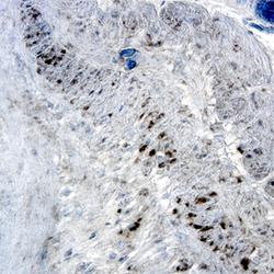
- Src in Human Colon. Src phosphorylated at Y419 was detected in immersion fixed paraffin-embedded sections of human colon using Rabbit Anti-Human Phospho-Src (Y419) Antigen Affinity-purified Polyclonal Antibody (Catalog # AF2685) at 15 µg/mL overnight at 4 °C. Tissue was stained using the Anti-Rabbit HRP-DAB Cell & Tissue Staining Kit (brown; Catalog # CTS005) and counterstained with hematoxylin (blue). Specific labeling was localized to the cytoplasm of epithelial cells. View our protocol for Chromogenic IHC Staining of Paraffin-embedded Tissue Sections.
