 Explore
 Validate
 Learn
Learn Western blot
Antibody data
- Antibody Data
- Antigen structure
- References [10]
- Comments [0]
- Validations
- Western blot [3]
- Immunohistochemistry [2]
- Other assay [1]
Submit
Validation data
Reference
Comment
Report error
- Product number
- AHO1152 - Provider product page

- Provider
- Invitrogen Antibodies
- Product name
- SRC Monoclonal Antibody (184Q20)
- Antibody type
- Monoclonal
- Antigen
- Recombinant full-length protein
- Reactivity
- Human, Mouse, Rat
- Host
- Mouse
- Isotype
- IgG
- Antibody clone number
- 184Q20
- Vial size
- 100 µg
- Concentration
- 0.5 mg/mL
- Storage
- -20°C
Submitted references Senescent hepatic stellate cells promote liver regeneration through IL-6 and ligands of CXCR2.
Differential Signaling Mediated by ApoE2, ApoE3, and ApoE4 in Human Neurons Parallels Alzheimer's Disease Risk.
Regulation of cytoskeleton and adhesion signaling in osteoclasts by tetraspanin CD82.
Treatment with Src inhibitor Dasatinib results in elevated metastatic potential in the 4T1 murine mammary carcinoma model.
Reelin signaling specifies the molecular identity of the pyramidal neuron distal dendritic compartment.
Impaired long term memory consolidation in transgenic mice overexpressing the human soluble form of IL-1ra in the brain.
Presenilin 1 affects focal adhesion site formation and cell force generation via c-Src transcriptional and posttranslational regulation.
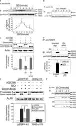
Beta-adrenergic receptor mediated protection against doxorubicin-induced apoptosis in cardiomyocytes: the impact of high ambient glucose.
Epidermal hyperplasia and papillomatosis in mice with a keratinocyte-restricted deletion of csk.
Analyzing FAK and Pyk2 in early integrin signaling events.
Cheng N, Kim KH, Lau LF
JCI insight 2022 Jun 16;7(14)
JCI insight 2022 Jun 16;7(14)
Differential Signaling Mediated by ApoE2, ApoE3, and ApoE4 in Human Neurons Parallels Alzheimer's Disease Risk.
Huang YA, Zhou B, Nabet AM, Wernig M, Südhof TC
The Journal of neuroscience : the official journal of the Society for Neuroscience 2019 Sep 11;39(37):7408-7427
The Journal of neuroscience : the official journal of the Society for Neuroscience 2019 Sep 11;39(37):7408-7427
Regulation of cytoskeleton and adhesion signaling in osteoclasts by tetraspanin CD82.
Bergsma A, Ganguly SS, Wiegand ME, Dick D, Williams BO, Miranti CK
Bone reports 2019 Jun;10:100196
Bone reports 2019 Jun;10:100196
Treatment with Src inhibitor Dasatinib results in elevated metastatic potential in the 4T1 murine mammary carcinoma model.
Hughes VS, Siemann DW
Tumor & microenvironment 2018;1(1):30-36
Tumor & microenvironment 2018;1(1):30-36
Reelin signaling specifies the molecular identity of the pyramidal neuron distal dendritic compartment.
Kupferman JV, Basu J, Russo MJ, Guevarra J, Cheung SK, Siegelbaum SA
Cell 2014 Sep 11;158(6):1335-1347
Cell 2014 Sep 11;158(6):1335-1347
Impaired long term memory consolidation in transgenic mice overexpressing the human soluble form of IL-1ra in the brain.
Spulber S, Mateos L, Oprica M, Cedazo-Minguez A, Bartfai T, Winblad B, Schultzberg M
Journal of neuroimmunology 2009 Mar 31;208(1-2):46-53
Journal of neuroimmunology 2009 Mar 31;208(1-2):46-53
Presenilin 1 affects focal adhesion site formation and cell force generation via c-Src transcriptional and posttranslational regulation.
Waschbüsch D, Born S, Niediek V, Kirchgessner N, Tamboli IY, Walter J, Merkel R, Hoffmann B
The Journal of biological chemistry 2009 Apr 10;284(15):10138-49
The Journal of biological chemistry 2009 Apr 10;284(15):10138-49
Beta-adrenergic receptor mediated protection against doxorubicin-induced apoptosis in cardiomyocytes: the impact of high ambient glucose.
Yano N, Suzuki D, Endoh M, Tseng A, Stabila JP, McGonnigal BG, Zhao TC, Padbury JF, Tseng YT
Endocrinology 2008 Dec;149(12):6449-61
Endocrinology 2008 Dec;149(12):6449-61
Epidermal hyperplasia and papillomatosis in mice with a keratinocyte-restricted deletion of csk.
Honda K, Sakaguchi T, Sakai K, Schmedt C, Ramirez A, Jorcano JL, Tarakhovsky A, Kamisoyama H, Sakai T
Carcinogenesis 2007 Oct;28(10):2074-81
Carcinogenesis 2007 Oct;28(10):2074-81
Analyzing FAK and Pyk2 in early integrin signaling events.
Bernard-Trifilo JA, Lim ST, Hou S, Schlaepfer DD, Ilic D
Current protocols in cell biology 2006 Apr;Chapter 14:Unit 14.7
Current protocols in cell biology 2006 Apr;Chapter 14:Unit 14.7
No comments: Submit comment
Supportive validation
- Submitted by
- Invitrogen Antibodies (provider)
- Main image
- Experimental details
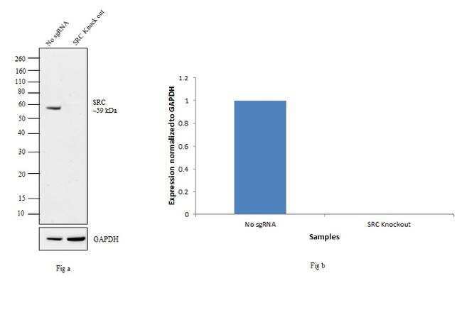
- Western blot analysis of SRC was performed by loading 30 µg of HeLa (Lane 1), HeLa- SRC knockout (Lane 2) whole cell lysate. The blot was probed with Anti-SRC Monoclonal Antibody (Product # AHO1152, 2 µg/mL) Goat anti-Mouse IgG (H+L) Superclonal™ Secondary Antibody, HRP conjugate (Product # A28177, 0.25 µg/mL 1:4000 dilution). Loss of signal upon CRISPR mediated knockout (KO) confirms that antibody is specific to SRC.
- Submitted by
- Invitrogen Antibodies (provider)
- Main image
- Experimental details
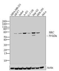
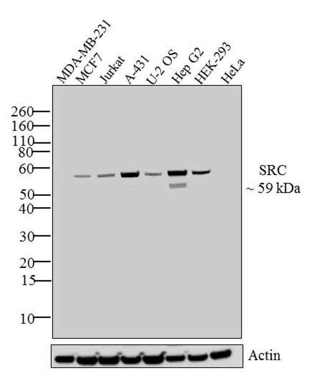
- Western blot analysis of SRC was performed by loading 30 µg of MDA-MB-231 (lane1), MCF 7 (lane2), Jurkat (lane3), A431 (lane4), U2OS (lane5), Hep G2 (lane 6), HEK-293 (lane 7) and HeLa (lane 8) cell lysate using Novex®NuPAGE®4-12 % Bis-Tris gel (Product # NP0322PK2), XCell SureLock Electrophoresis System (Product # EI0002), Novex® Sharp Pre-Stained Protein Standard (LC5800), and iBlot® Dry Blotting System (IB21001). Proteins were transferred to a nitrocellulose membrane and blocked with 5% skim milk for 1 hour at room temperature. SRC was detected at ~ 59 kDa using SRC Mouse Monoclonal Antibody (Product # AHO1152) at 1-3 µg/mL in 5% skim milk at 4°C overnight on a rocking platform. Goat Anti-Mouse IgG - HRP Secondary Antibody (Product # 62-6520) at 1:4000 dilution was used and chemiluminescent detection was performed using Pierce™ ECL Western Blotting Substrate (Product # 32106).
- Submitted by
- Invitrogen Antibodies (provider)
- Main image
- Experimental details
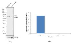
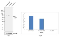
- Knockdown of SRC was achieved by transfecting A-431 cells with SRC specific siRNAs (Silencer® select Product # s13414). Western blot analysis (Fig a) was performed using membrane extracts from the SRC knock down cells (lane 3), non-specific scrambled siRNA transfected cells (lane 2) and untransfected cells (lane 1). The blots were probed with Anti-SRC Mouse monoclonal Antibody (Product # AHO1152, 2 µg/mL) and Goat anti-Mouse IgG (H+L) Superclonal™ Secondary Antibody, HRP conjugate (Product # A28177, 0.4 µg/mL, 1:5000 dilution). Densitometric analysis of this western blot is shown in histogram (Fig b). Loss of signal upon siRNA mediated knock down confirms that antibody is specific to SRC.

Supportive validation
- Submitted by
- Invitrogen Antibodies (provider)
- Main image
- Experimental details
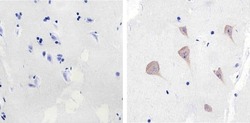
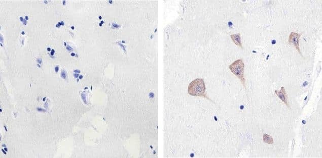
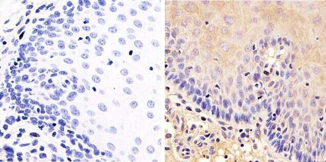
- Immunohistochemistry analysis of SRC showing staining in the cytoplasm of paraffin-embedded human brain tissue (right) compared to a negative control without primary antibody (left). To expose target proteins, antigen retrieval was performed using 10mM sodium citrate (pH 6.0), microwaved for 8-15 min. Following antigen retrieval, tissues were blocked in 3% H2O2-methanol for 15 min at room temperature, washed with ddH2O and PBS, and then probed with a SRC (Product # AHO1152) diluted in 3% BSA-PBS at a dilution of 1:100 overnight at 4°C in a humidified chamber. Tissues were washed extensively in PBST and detection was performed using an HRP-conjugated secondary antibody followed by colorimetric detection using a DAB kit. Tissues were counterstained with hematoxylin and dehydrated with ethanol and xylene to prep for mounting.
- Submitted by
- Invitrogen Antibodies (provider)
- Main image
- Experimental details
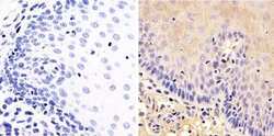
- Immunohistochemistry analysis of SRC showing staining in the cytoplasm of paraffin-embedded human esophagus tissue (right) compared to a negative control without primary antibody (left). To expose target proteins, antigen retrieval was performed using 10mM sodium citrate (pH 6.0), microwaved for 8-15 min. Following antigen retrieval, tissues were blocked in 3% H2O2-methanol for 15 min at room temperature, washed with ddH2O and PBS, and then probed with a SRC (Product # AHO1152) diluted in 3% BSA-PBS at a dilution of 1:100 overnight at 4°C in a humidified chamber. Tissues were washed extensively in PBST and detection was performed using an HRP-conjugated secondary antibody followed by colorimetric detection using a DAB kit. Tissues were counterstained with hematoxylin and dehydrated with ethanol and xylene to prep for mounting.
Supportive validation
- Submitted by
- Invitrogen Antibodies (provider)
- Main image
- Experimental details
- NULL
